Abstract
Athletes usually complain of an ongoing or chronic pain over the Achilles tendon, but recently even non-athletes are experiencing the same kind of pain which affects their daily activities. Achilles tendinosis refers to a degenerative process of the tendon without histologic or clinical signs of intratendinous inflammation. Treatment is based on whether to stimulate or prevent neovascularization. Thus, until now, there is no consensus as to the best treatment for this condition. This paper aims to review the common ways of treating this condition from the conservative to the surgical options.
Professional and recreational athletes, as well as, sedentary people often complain of tendon pain. Most tendon injuries are the result of gradual wear and tear from overuse or aging. Anyone can have a tendon injury but people who make repetitive movements in their jobs, sports, or daily activities are more likely to damage a tendon.
Achilles tendinopathy is a painful condition that occurs commonly in active and inactive individuals.1) It was initially reported to be a tendon disorder which has multiple suggested pathology which are based on poor scientific evidence as explained by Lake and Ishikawa.1) But later researches have clarified the difference between an Achilles tendinitis and an Achilles tendinosis. An Achilles tendinitis (tendonitis) occurs when there is a clinical presence of pain and swelling. It is an inflammatory process seen on a biopsy specimen of a diseased tendon. On the other hand, an Achilles tendinosis refers to a degenerative process of the tendon without histologic or clinical signs of intratendinous inflammation. Leadbetter2) suggested that tendinosis is a failure of the cell matrix to adapt to repetitive trauma caused by an imbalance between the degeneration and synthesis of the matrix. An isolated pain at the insertion of the Achilles tendon to the calcaneus due to an intratendinous degeneration is referred to as insertional Achilles tendinosis, while a non-insertional (mid-portion) Achilles tendinosis occurs in the main body of the Achilles tendon.
The etiology behind an Achilles tendinosis remains unclear but there are many theories as to the cause of the disease which include overuse, decreased blood supply and tensile strength with aging, muscle imbalance or weakness, insufficient flexibility, and even malalignment such as hyperpronation.1) Some also suggest that genetics, endocrine disorders, and free-radical production can also produce tendinosis. Biopsies of diseased tendons revealed that there are cellular activation evidenced by an increase in cell numbers and ground substance, collagen disarray, and neovascularisation. Prostaglandin inflammatory elements are not present but neurogenic elements such as substance-P and calcitonin gene-related peptides have been isolated.1) Neurovascular in-growth and glutamate (a potent modulator of pain) has been noted in the diseased tendons and they have been postulated to be the sources of pain in patients with Achilles tendinosis.
Tendon injuries occur in 30%-50% of all sports related injuries. Sixty-six percent of joggers complain of Achilles tendon pain and 23% of them usually have insertional Achilles tendinosis.3) Risk factors for the development of this condition include diabetes, hypertension, obesity, hormone replacement, and use of oral contraceptives.4) One study even suggests that having a pes cavus could also cause Achilles tendinosis. Since there is no gold standard treatment for the Achilles tendinosis, this paper aims to describe the various treatment options for the disorder, conservative as well as surgical treatments.
The first line of treatment for any kind of disease is still the non-invasive methods such as activity modification, orthotics, heel lifts, massage, hot and cold compresses, strengthening exercises, ultrasound, and non-steroidal anti-inflammatory drugs (NSAID) or oral corticosteroid.3) Since there are no prostaglandin inflammatory mediators in an Achilles tendinosis, NSAIDs has been questioned with regards to its effectiveness.5) In a randomized double-blind placebo-controlled trial done by Astrom and Westlin,5) it was concluded that piroxicam was no more effective than placebo. Though the traditional non-invasive methods are readily available and give most of the patients short-term pain relief, 30% of them still find these alternatives as ineffective.
Corticosteroids are a class of medications that are related to a steroid called cortisone. They relieve pain by reducing the inflammation that occurs in a diseased tendon. There have been many conflicting studies regarding the use of corticosteroid injections in Achilles tendinosis since there is no inflammatory process. It produces short-term pain relief after peritendinous corticosteroid injection in one randomized control6) trial but another7) demonstrated no improvement. Gill et al.8) reported 40% improvement without tendon rupture. Since their role in Achilles tendinosis has not yet been established, precaution is always advised as there have been multiple reports of complete or partial ruptures of the Achilles tendon.1)
The eccentric training program developed by Alfredson et al.9) promotes tendon healing by increasing the tendon volume and signal intensity which is thought to be a response to trauma. But after a 12-week program, a decrease in size and a more normal tendon appearance were noted on ultrasound and magnetic resonance imaging (MRI).10) With continuous eccentric loading of the Achilles tendon, there will be lengthening of the muscle-tendon unit and increase the tendons capacity to bear load overtime. Repetitive eccentric training may cause damage to the abnormal blood vessel and the accompanying nerves in the tendon thus eliminating the pain as concluded by Alfredson et al.11) With the 12-week program, it has produced 90% good results with mid-portion Achilles tendinosis and 30% good results in insertional Achilles tendinosis.1) In another study, the program produced 28% good results in insertional Achilles tendinosis.12)
The extracorporeal shock wave therapy (ESWT) is another treatment option for Achilles tendinosis. It can be given as a low energy treatment (which is 3 weekly sessions without local anesthesia or intravenous anesthesia) or as a high energy treatment (which is a single session but requires local or intravenous anesthesia). Microtrauma is produced by the repeated shock wave to the affected area which then stimulates neovascularization (Fig. 1). It is this new blood flow that promotes tissue healing and relief from pain. It can also inhibit afferent pain receptor function and produce a high number of nitric oxide synthase. In two non-randomized clinical trials, Furia13,14) reported that patients treated with high energy ESWT had more successful results than those treated with other traditional nonoperative treatment. This result is in contrast to Costa and colleagues two double-blinded, randomized controlled trial where they reported no statistically significant treatment effects in 49 patients with Achilles tendinosis treated with low-energy ESWT.1) Though not yet approved by the Food and Drug Administration for the treatment of Achilles tendinosis and the lack of availability of the machines, this treatment option seems to be beneficial and may have a place in the treatment of Achilles tendinosis.1)
Polidocanol is a sclerosing agent used to sclerose neovascularization. It causes thrombosis through a selective effect on the intima even when the drug is injected extravascularly. Based from European literature, when the process of neovascularization in the injured tendon is eliminated, the new blood vessels including the sensory nerves that are linked with them are destroyed producing pain relief to the patient.1) Protocol states that the sclerosing agent is injected 2-3 times with 6-8 weeks apart.11) Each injection is followed by a few days of rest, and high-impact activities are restricted for 2 weeks. Alfredson et al.11) demonstrated neurovascular in-growth in painful tendons and a reduction in pain with sclerosis of the neo-vessels in both the patellar and Achilles tendons. However, there have been reports of tendon ruptures in elite athletes after multiple sclerosant injections.1)
Sclerosing thermal therapy uses a radiofrequency probe to carry out microtenotomies. Similar to sclerosing agents, the thermal energy applied to the diseased tendon destroys the new blood vessels together with the sensory nerves that accompany them. Boesen et al.15) demonstrated good results after using an ultrasound-guided electro-cauterization technique in 11 patients with chronic mid-portion tendinosis. The result of the study showed that 10 out of 11 patients had 0 level of pain from a median score of 7 using the Likert box scale after six months of only one treatment session. One patient dropped out from the study due to disappointment after receiving an additional treatment without improvement.
Glyceryltrinitrate is a prodrug of endogenous nitric oxide. It is commercially available as a topical patch to relieve nitric oxide which is a soluble gas that acts as a messenger molecule that can affect many cellular functions, including tendon healing.16) It is believed that it increases collagen production by fibroblasts, cellular adhesion, and local vascularity. However, a study by Osadnik et al.17) concluded that a topical glyceryltrinitrate facilitates capillary venous outflow in painful Achilles tendons and that the capillary blood flow and tendon oxygenation remain unchanged. In a randomized double-blind placebo-controlled study by Hunte and Lloyd-Smith,18) a topical glyceryltrinitrate patch was more effective than placebo for reducing pain from chronic non-insertional Achilles tendinosis in the first 12 and 24 weeks of use. Another study showed excellent results (78%) for Achilles tendons treated with 1.25 mg. of topical glyceryltrinitrate every 24 hours for 6 months as compared to the placebo group (49%). A follow-up of the same study was done after 3 years and showed that 88% of the patients treated with topical glyceryltrinitrate and 67% of placebo patients were asymptomatic.19) Kane et al.20) however concluded that there were no difference with the Ankle Osteoarthritis Scale visual analog score for pain and disability between those treated with topical glyceryltrinitrate and those who underwent formal physical therapy program for 6 months. Therefore, there are still conflicting reports about the use of this medication and further evaluation is still recommended.
Aprotinin is a serine protease inhibitor. It is a strong inhibitor of matrix metalloproteinases which are found to be increased in diseased tendons. Its main effect is slowing down of fibrinolysis, the process that leads to the breakdown of blood clots. This is done as a one-time dose or a delay of at least 6 weeks between dosing to lower the risk of severe allergic reaction, as there had been reports of pruritus due to anaphylaxis (3%-11%).21) A success rate of 80% in Achilles tendinosis has been reported in one uncontrolled study.22)
Low level laser therapy produces effects on a diseased tendon like enhanced adenosine triphosphate production, enhanced cell function, and increased protein synthesis.23) It also reduces inflammation, increases collagen synthesis, and promotes angiogenesis. In a study done by Stergioulas et al.,23) they concluded that Achilles tendinosis patients who underwent eccentric exercises together with low level laser therapy showed decreased pain intensity, morning stiffness, tenderness to palpation, active dorsiflexion, and crepitus with no side effects as compared to those who underwent eccentric exercises only. However, there is still limited data to verify the effectiveness of low level laser therapy for the treatment of Achilles tendinosis.
This is a series of a hypertonic glucose with lignocaine/lidocaine injection designed to sclerose the new blood vessels and nerves. A randomized control trial done by Sweeting and Yelland24) compared the outcomes of eccentric loading exercises and prolotherapy injections used singly and in combination for non-insertional Achilles tendinosis. It showed that as early as 6 weeks, the combination therapy produced more rapid improvements with regards to symptoms of pain compared to monotherapy in selected subjects.
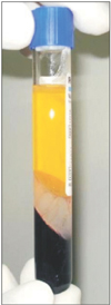
In chronic Achilles tendinosis, there is an absence of inflammation and a paucity of platelets.25) Platelets when activated produce cytokines and granules that then produce growth factors that aid in the healing process.25) Increasing the concentration of platelets through injection to the tissue enhances the healing potential by stimulating revascularization of the injured tendon. There are no established indications for the use of platelet rich plasma (PRP) in Achilles tendinosis although the current best evidence suggests that patients will improve after PRP treatment (Fig. 2). However, the improvement is not significantly better than physical therapy. de Jonge et al.26) with a randomized control trial concluded that there were no clinical and ultrasonographic superiority of PRP injection over a placebo injection in chronic Achilles tendinosis at one year combined with an eccentric training program.
Operative treatment of Achilles tendinosis involves the removal of abnormal tissues and lesions, fenestration of the tendon through multiple longitudinal creations, and possibly stripping the paratenon. The goal of such management is to remove degenerative nodules, excise fibrotic adhesions, restore the vascularity, and stimulate viable cells to initiate an inflammatory response and reinitiate healing. Results showed that an open surgical treatment for an Achilles tendinosis produced 18.8% unsatisfactory outcome in non-athletic subjects as compared to athletic subjects (8.9%).27) A study also showed that reoperation rate is higher in women (12.2%) as compared to men (6.7%).28)
Testa et al.29) developed a technique where multiple, ultrasound-guided, percutaneous incisions are made through the diseased Achilles tendon. This procedure can be done as an out-patient surgery where the patient is placed on prone position. Local anesthesia is applied over the diseased area which can be identified through palpation or ultrasound. A stab knife is then used to make a longitudinal incision parallel to the long axis of the Achilles tendon. With the knife pointing cephalad, the ankle is then fully dorsiflexed. Then with the scalpel pointing towards the caudal direction, the ankle is then fully plantarflexed. Four separate stab incisions are made approximately 2 cm apart; 1 medial and proximal, 1 medial and distal, 1 lateral and proximal, and 1 lateral and distal. The incisions are closed with adhesive strips. Early range of motion is encouraged and full weight bearing is allowed after 2-3 days with an expected return to previous activity after 4-6 weeks. Testa et al.29) and Maffulli et al.30) achieved 56% excellent results with this treatment and only 8% poor results. Maffulli et al.31) modified this technique in 2009 by adding another stab wound at the central portion of the diseased area (Fig. 3).
Longo et al.32) introduced a technique of stripping the adhesions in the Achilles tendon through a minimally invasive technique in 2008. This involves four 0.5 cm longitudinal skin incisions along the border of the Achilles tendon. Two are made just medial and lateral to the origin of the tendon and two more at distal end of the tendon close to the insertion. A mosquito is then inserted through the incisions and the proximal and distal portions of the Achilles tendon are freed of peritendinous adhesions. A number 1 Ethibond thread is inserted at the 2 proximal incisions over the anterior aspect of the Achilles tendon. Ends of the Ethibond are then retrieved from the distal incisions. The Ethibond is then slid onto the tendon, causing it to be stripped and freed from adhesions at the anterior surface of the tendon. The procedure is repeated for the posterior aspect of the tendon. This will disrupt the neovascularisation of the damaged tendon and its accompanying nerve supply. After the procedure, the patient is allowed to do range of motion exercises and can be allowed to do full weight-bearing.31)
Small skin incisions are made and an arthroscopic shaver is introduced into the Achilles tendon to debris the peritenon. This procedure produces decreased postoperative complication thus allowing the patient early return to previous activity.33) Steenstra and van Dijk33) reported significant pain relief after 2-7 years of 20 patients who were able to return to sports after 4-6 weeks. Maquirriain in a long term follow up study (5 years) reported a high rate of excellent results in patients with chronic Achilles tendinosis with 0% infection and systemic complications.34) There was however a report of delayed keloid lesion and a seroma with chronic fistula formation in his study postoperatively.
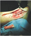
This procedure is suggested by some to be used for moderate to severe tendinosis or when conservative measures have already been exhausted. Contraindications are minimal preoperative pain or skin and vascular compromise. The paratenon is carefully incised and any inflammatory peritendinits is removed. If on MRI or ultrasound there is an intratendinous nodule, or there is a palpable thickening within the tendon, excision is recommended until viable tissues are seen.35) Any residual degenerated tissue increases the risk of persistent postoperative pain.35) If there is a Haglund's deformity, it can also be excised at this point (Fig. 4). A turned-down tendon flap may be necessary in order to bridge the gap left by the excision. The flexor hallucis longus can be used to augment the Achilles tendon by tendon transfer if more than 50% of diseased tendon was removed (Fig. 5). Schon et al.36) reported significant improvement in terms of Achilles tendon function, physical function and pain intensity with this procedure in relatively inactive, older, and overweight patients. However, there are some reports of tendon rupture after an open debridement.
There are a variety of conservative and surgical treatment options for an Achilles tendinosis, which implies the absence of the gold standard of the treatments. Mechanism of action for the ideal treatment is still controversial. Conservative measures such as corticosteroid injection reduces inflammation, while eccentric training lengthens muscle-tendon unit. ESWT or PRP stimulates neovascularization while the prolotherapy are used to sclerose neovascularization. Surgical measures include tendon micro-tenotomy, tendon debridement and paratenon stripping.
Figures and Tables
References
1. Lake JE, Ishikawa SN. Conservative treatment of Achilles tendinopathy: emerging techniques. Foot Ankle Clin. 2009; 14(4):663–674.
2. Leadbetter WB. Cell-matrix response in tendon injury. Clin Sports Med. 1992; 11(3):533–578.
3. Kvist M. Achilles tendon injuries in athletes. Sports Med. 1994; 18(3):173–201.
4. Holmes GB, Lin J. Etiologic factors associated with symptomatic Achilles tendinopathy. Foot Ankle Int. 2006; 27(11):952–959.
5. Astrom M, Westlin N. No effect of piroxicam on Achilles tendinopathy: a randomized study of 70 patients. Acta Orthop Scand. 1992; 63(6):631–634.
6. Smidt N, van der Windt DA, Assendelft WJ, Deville WL, Korthals-de Bos IB, Bouter LM. Corticosteroid injections, physiotherapy, or a wait-and-see policy for lateral epicondylitis: a randomised controlled trial. Lancet. 2002; 359(9307):657–662.
7. DaCruz DJ, Geeson M, Allen MJ, Phair I. Achilles paratendonitis: an evaluation of steroid injection. Br J Sports Med. 1988; 22(2):64–65.
8. Gill SS, Gelbke MK, Mattson SL, Anderson MW, Hurwitz SR. Fluoroscopically guided low-volume peritendinous corticosteroid injection for Achilles tendinopathy: a safety study. J Bone Joint Surg Am. 2004; 86(4):802–806.
9. Alfredson H, Pietila T, Jonsson P, Lorentzon R. Heavy-load eccentric calf muscle training for the treatment of chronic Achilles tendinosis. Am J Sports Med. 1998; 26(3):360–366.
10. Shalabi A, Kristoffersen-Wiberg M, Aspelin P, Movin T. Immediate Achilles tendon response after strength training evaluated by MRI. Med Sci Sports Exerc. 2004; 36(11):1841–1846.
11. Alfredson H, Ohberg L, Forsgren S. Is vasculo-neural ingrowth the cause of pain in chronic Achilles tendinosis? An investigation using ultrasonography and colour Doppler, immunohistochemistry, and diagnostic injections. Knee Surg Sports Traumatol Arthrosc. 2003; 11(5):334–338.
12. Fahlstrom M, Jonsson P, Lorentzon R, Alfredson H. Chronic Achilles tendon pain treated with eccentric calf-muscle training. Knee Surg Sports Traumatol Arthrosc. 2003; 11(5):327–333.
13. Furia JP. High-energy extracorporeal shock wave therapy as a treatment for insertional Achilles tendinopathy. Am J Sports Med. 2006; 34(5):733–740.
14. Furia JP. High-energy extracorporeal shock wave therapy as a treatment for chronic noninsertional Achilles tendinopathy. Am J Sports Med. 2008; 36(3):502–508.
15. Boesen MI, Torp-Pedersen S, Koenig MJ, et al. Ultrasound guided electrocoagulation in patients with chronic non-insertional Achilles tendinopathy: a pilot study. Br J Sports Med. 2006; 40(9):761–766.
16. Murrell GA. Using nitric oxide to treat tendinopathy. Br J Sports Med. 2007; 41(4):227–231.
17. Osadnik R, Redeker J, Kraemer R, Vogt PM, Knobloch K. Microcirculatory effects of topical glyceryl trinitrate on the Achilles tendon microcirculation in patients with previous Achilles tendon rupture. Knee Surg Sports Traumatol Arthrosc. 2010; 18(7):977–981.
18. Hunte G, Lloyd-Smith R. Topical glyceryl trinitrate for chronic Achilles tendinopathy. Clin J Sport Med. 2005; 15(2):116–117.
19. Paoloni JA, Murrell GA. Three-year followup study of topical glyceryl trinitrate treatment of chronic noninsertional Achilles tendinopathy. Foot Ankle Int. 2007; 28(10):1064–1068.
20. Kane TP, Ismail M, Calder JD. Topical glyceryl trinitrate and noninsertional Achilles tendinopathy: a clinical and cellular investigation. Am J Sports Med. 2008; 36(6):1160–1163.
21. Orchard J, Massey A, Rimmer J, Hofman J, Brown R. Delay of 6 weeks between aprotinin injections for tendinopathy reduces risk of allergic reaction. J Sci Med Sport. 2008; 11(5):473–480.
22. Capasso G, Maffulli N, Testa V, Sgambato A. Preliminary results with peritendinous protease inhibitor injections in the management of Achilles tendinitis. J Sports Traumatol Rel Res. 1993; 15(1):37–43.
23. Stergioulas A, Stergioula M, Aarskog R, Lopes-Martins RA, Bjordal JM. Effects of low-level laser therapy and eccentric exercises in the treatment of recreational athletes with chronic achilles tendinopathy. Am J Sports Med. 2008; 36(5):881–887.
24. Sweeting K, Yelland M. Achilles tendinosis: how does prolotherapy compare to eccentric loading exercises? J Sci Med Sport. 2009; 12:Supplement. S19.
25. Magnussen RA, Dunn WR, Thomson AB. Nonoperative treatment of midportion Achilles tendinopathy: a systematic review. Clin J Sport Med. 2009; 19(1):54–64.
26. de Jonge S, de Vos RJ, Weir A, et al. One-year follow-up of platelet-rich plasma treatment in chronic Achilles tendinopathy: a double-blind randomized placebo-controlled trial. Am J Sports Med. 2011; 39(8):1623–1629.
27. Maffulli N, Testa V, Capasso G, et al. Surgery for chronic Achilles tendinopathy yields worse results in nonathletic patients. Clin J Sport Med. 2006; 16(2):123–128.
28. Maffulli N, Testa V, Capasso G, et al. Surgery for chronic Achilles tendinopathy produces worse results in women. Disabil Rehabil. 2008; 30(20-22):1714–1720.
29. Testa V, Capasso G, Benazzo F, Maffulli N. Management of Achilles tendinopathy by ultrasound-guided percutaneous tenotomy. Med Sci Sports Exerc. 2002; 34(4):573–580.
30. Maffulli N, Testa V, Capasso G, Bifulco G, Binfield PM. Results of percutaneous longitudinal tenotomy for Achilles tendinopathy in middle- and long-distance runners. Am J Sports Med. 1997; 25(6):835–840.
31. Maffulli N, Longo UG, Spiezia F, Denaro V. Minimally invasive surgery for Achilles tendon pathologies. Open Access J Sports Med. 2010; 1:95–103.
32. Longo UG, Ramamurthy C, Denaro V, Maffulli N. Minimally invasive stripping for chronic Achilles tendinopathy. Disabil Rehabil. 2008; 30(20-22):1709–1713.
33. Steenstra F, van Dijk CN. Achilles tendoscopy. Foot Ankle Clin. 2006; 11(2):429–438.
34. Maquirriain J. Surgical treatment of chronic achilles tendinopathy: long-term results of the endoscopic technique. J Foot Ankle Surg. 2013; 52(4):451–455.
35. Murphy GA. Surgical treatment of non-insertional Achilles tendinitis. Foot Ankle Clin. 2009; 14(4):651–661.
36. Schon LC, Shores JL, Faro FD, Vora AM, Camire LM, Guyton GP. Flexor hallucis longus tendon transfer in treatment of Achilles tendinosis. J Bone Joint Surg Am. 2013; 95(1):54–60.


 PDF
PDF ePub
ePub Citation
 Print
Print


 XML Download