

 PDF
PDF ePub
ePub Citation
 Print
Print


Heterotrophic ossification of the upper limb is a rare post-traumatic complication.1) Moreover, it is not very common among children. The localized form of myositis ossificans of the upper limb often occurs in the brachialis muscle. When such ossification occurs around a joint, fixed deformities and complete restriction of motion can result. Although the condition is said to be self limiting,2) excision is unavoidable in circumstances where the myositic mass limits daily activities. High suspicion is needed to diagnose this condition clinically. A painless enlarging mass occurring after single or repeated trauma should lead to a suspicion of myositis ossificans.3) Ultrasound or radiographs should be used to make the clinicoradiological diagnosis. We experienced an unusually large myositic mass in a teenage boy, which restricted elbow motion. Complete surgical excision led to significant improvement of elbow function.
CASE REPORT
A 13-year-old boy presented with a 14-month history of a fixed left elbow with no movement. He had sustained significant trauma to the affected limb 1 month prior to onset of symptoms. He had received treatment in the form of native massage and bandaging with immobilization for 4 weeks. However, he had difficulty moving his left elbow, which resulted in a fixed flexion deformity with no further movement. He had difficulties with daily activities such as clothing himself and tying shoes. However, he managed using the normal limb, as he was right handed.
A clinical examination revealed bony ankylosis of the left elbow at 40° of flexion. A palpable diffuse irregular bony mass was present in the anterior aspect of the elbow but the margins could not be delineated. No part of the bony mass or any other bony prominence was tender and warm. The arm and the forearm segment appeared to be wasted when compared to those of the other limb. No distal neurovascular compromise or any similar lesion was detected elsewhere in the body.
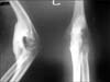
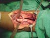
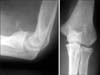
Standard anteroposterior and lateral radiographs of the affected elbow revealed an unusually large mass connecting the distal humerus to the ulna (Fig. 1). Radio-opacity of the mass was similar to that of mature bone. Complete hemogram and erythrocyte sedimentation rate values were within normal ranges. Considering the physical demand of the patient and the unusual size of the myositic mass, surgical excision was planned. The procedure was performed under regional anesthesia with the patient in the supine position and the limb abducted at the shoulder. An 8-cm curvilinear incision starting about 3 cm proximal to the elbow crease and extending to the junction of the proximal and mid-third of the forearm was made. Skin, subcutaneous tissue, and deep fascia were incised in line with the skin incision. The neurovascular bundle was identified and retracted. The extent of the abnormal bone was exposed adequately (Fig. 2).
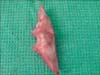
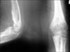
The mass was excised completely after detaching it from the humerus and ulna (Fig. 3). The elbow was mobilized intraoperatively, and full range resumed. The excised specimen was sent for histopathological examination and revealed lamellar bone both peripherally and centrally. The child developed neuropraxia of the median nerve postoperatively, which recovered within 2 weeks. Postoperative radiographs showed no trace of the myositic mass (Fig. 4). Elbow range of motion exercises were started by the middle of the first week, as tolerated. Active flexion of 30° to 90° was possible during the initial postoperative period. The patient was reviewed every 2 weeks for the first 2 months and once monthly for up to 6 months to assess functional status.
The patient regained full function of the affected elbow and returned to school in 10 weeks. He scored the maximum of 100 on the Mayo Elbow Scoring System, which was attributed to full functional status. The patient had the best possible functional status after 2 years. No signs of clinical or radiographic recurrence were detected (Fig. 5).
DISCUSSION
Myositis ossificans traumatica of the upper limb causing ankylosis of the elbow is a rare entity. The specific cause for myositis and its pathophysiology remain unclear. Proliferation of connective tissue occurs after a muscle injury that differentiates into mature bone. Such ossification preferably occurs in muscles that are repeatedly strained or injuried.3) The condition is said to be self limiting, and spontaneous resolution occurs over a period of time.2) Lesions result in significant functional deficit in only 10%-20% of patients.4) The most commonly involved regions are the thighs, hips, upper arms, calves, and soles of the feet.5,6) Myositis ossificans has also been reported at some other rare sites.7,8) Native bandaging and repeated massage for traumatic conditions are still common in some parts of Asia. This treatment predisposes to myositis, particularly around the elbow in children. Small specks of calcification usually occur either anterior or posterior. However, the mass connecting the humerus to the ulna was almost the same diameter as the humerus in this case.
The early clinical course of myositis ossificans traumatica closely resembles that of osteosarcoma with localized swelling and tenderness. Pain subsides after ossification, unlike osteosarcoma. Radiological and histopathological findings can satisfactorily distinguish between the two. Radiographs of osteosarcoma show periosteal elevation and cortical destruction, which are not present in myositis ossificans. During the early stage, computerized tomography demonstrates a zoning phenomenon in a myositis mass. Ultrasound also shows a center of less echogenicity with an outer sheet-like hyperechoic peripheral rim. The late stage radiographically resembles fully ossified bone.
A histopathological analysis reveals a variable zonal pattern with a central zone of rapidly growing fibroblasts, an intermediate zone of osteoblasts with immature osteoids, and a peripheral zone of mature bone. The late stage shows a well organized cortex and a marrow space resembling mature bone.9) Conservative management with physical therapy, acetic acid iontophoresis, magnesium therapy, and etidronate disodium are reported to be effective.10) Surgical excision is indicated only if the lesion is completely ossified because premature excision leads to local recurrence.
Our patient had fully mature bone bridging the distal humerus to the proximal ulna at presentation. Surgical excision led to significant improvement and limited the disability, which was the major concern. Early postoperative mobilization and physiotherapy helped our patient regain near normal function of the affected limb. The patient scored the maximum number of points on the Mayo Elbow Scoring System and recovered full function of the affected elbow. A 2-year follow-up revealed no signs of recurrence and full power in the elbow.
Myositis ossificans traumatica is one of the most debilitating complications of muscle contusion and can result in gross limitations in joint function. Many children who have received native treatment and massage present with myositis, particularly of the elbow. We commonly observe small calcification specks. The acute stage results in immobilization. Chronic myositis may present as a small globular mass in the brachialis muscle. However, in our case, the mass was of unusual size and was ankylosing the joint. Surgical excision produced a satisfactory outcome.
 XML Download