

 PDF
PDF ePub
ePub Citation
 Print
Print


Arthrodesis of the distal interphalangeal (DIP) joint of the hand is commonly performed to treat a variety of conditions including pain, deformity, and instability. These problems usually derive from degenerative arthritis, inflammatory diseases or traumatic problems such as irreparable damage to the relevant flexor or extensor tendon, and joint destruction. Stable bony union is essential for successful arthrodesis, and discomfort caused by the hardware should be absent. Additionally, early joint movement should be allowed to prevent adjacent joint stiffness. Arthrodesis of the DIP joints has a long history as evidenced by the variety of developed techniques, including crossed Kirschner wires (K-wires), screw or plate fixation and external fixation.1,2,3,4) Although results from multiple techniques of DIP joint arthrodesis are often very promising, each procedure has some degree of difficulty or requires specific instruments.1,2,3) We applied a simple procedure of intramedullary K-wire fixation and interosseous wiring. The purpose of this study was to introduce our technique of DIP joint arthrodesis using simple instruments and to assess its objective and subjective outcomes.
METHODS
We retrospectively reviewed a group of patients who had undergone arthrodesis of the DIP joints, using implantable intramedullay K-wire fixation and interosseous wiring, from October 2008 and March 2012. The study included 9 patients (7 women and 2 men) and the mean age of patients was 44.2 years (range, 21 to 71 years). Painful osteoarthritis was observed in 4 patients, chronic irreparable mallet finger in 3 patients and post-traumatic arthritis in 2 patients. The dominant hand was affected in 6 patients, and the affected fingers were 4 ring fingers, 3 long fingers and 1 index and small finger each.
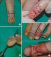
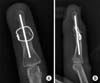
Surgical procedures were performed under local regional anesthesia through an H-shaped mid-dorsal skin incision centering over the DIP joint (Fig. 1A). The extensor tendon and the capsule were transected to expose the joint and the collateral ligaments were released. After removing the synovium and osteophyte, the articular surface of the distal phalanx base and the middle phalanx head were resected with an electric saw, creating cancellous surfaces perpendicular to the longitudinal axes of both phalanges. Thickness of resection was less than 2 mm in each surface and we tried to keep the total amount of shortening to less than 4 mm for maintaining finger length. A 1.1-mm K-wire was drilled anterogradely through the base of the distal phalanx and penetrated to the outside of the finger tip for preparation of intramedullary holes. The K-wire was then withdrawn and reinserted retrogradely into the medullary canal of the midphalanx across the joint (Fig. 1B). The entire length of K-wire was positioned inside the medullary space by cutting the distal part and further extended into the middle phalanx canal (Fig. 1C). Eighteen-gauge intravenous catheter needles were inserted by bicortical transverse drilling across the neck of the middle phalanx and the base of the distal phalanx for passing the interosseous wire (Fig. 1D). A 26-gauge steel wire was passed inside the needles and the needles were then removed. Contact surface was compressed and secure fixation was achieved by tensioning of the interosseous wire (Fig. 1E) with straight arthrodesis position. Positioning of the K-wire and the interosseous wire was verified by fluoroscopy at the end of the procedure (Fig. 2). A short aluminum splint was applied for protection of the DIP joint and full range of motion exercises of the proximal interphalangeal and metacarpophalangeal joints were encouraged immediately after surgery. The splint for DIP joint was removed postoperative 4 weeks and gradual pinch movement was allowed from 6 weeks after surgery.
Radiologic union was evaluated through the anteroposterior and lateral views of the radiographs which were checked every 2 weeks, until evidence of union. Union was defined by the presence of bony trabeculae crossing the arthrodesis site. Visual analogue scale (VAS) of the DIP joint was recorded to evaluate the remaining pain postoperatively. We also investigated the presence of any immediate of delayed surgical complications.
RESULTS
The mean follow-up period was 19.6 months (range, 12 to 36 months) and all cases had successful fusion of the joint. Patients showed union of the joint on radiographs taken from 6 to 10 weeks after surgery, and the mean time was 7.6 weeks. Average VAS score was improved from 6.5 points preoperatively to 1.2 points (range, 0 to 3 points) at the last follow-up in arthritis patients. Cases of chronic mallet finger were excluded from VAS score evaluation because their main complaint was deformity without painful arthritic change. We noted no malunion, nail deformity, hardware migration or infection on physical examination or in follow-up radiographs. Wire removal was required in 1 patient, due to irritation by the knot of the interosseous wire at 4 months postoperatively and satisfactory relief of discomfort was achieved during the follow-up.
DISCUSSION
Arthrodesis is a generally accepted operative treatment for arthritis of the DIP joint to relieve pain and to correct deformity and instability in patients who are unresponsive to conservative measures. Bunnell introduced a technique for arthrodesis consisting of resection of the articular surfaces and securing of the joint with two K-wires. Since then, many similar techniques have been introduced.5,6) Because each technique has its own advantages and problems, no single technique has gained universal popularity.1)
K-wires have been used for many years, leading to insufficient osseous stabilization7) and making it necessary to remove the K-wire after the union. A correlation was found between infection and the use of K-wires in another study.8) We observed no postoperative complication with suspected infection. Our opinion was that the intramedullary location of the entire length of K-wire lowers the risk of infection from exposed K-wire tip outside the skin. More recently, headless compression screw or plate fixation for interphalangeal joint fusion has demonstrated consistent success rates.1,3) Villani et al.3) reported 102 cases of headless compression screw fixation for DIP joint arthrodesis, with 2 cases of prominent hardware, 1 complex regional pain syndrome and 1 symptomatic bony callus from 89 fused joints. Mantovani et al.2) also reported a 100% union rate of the DIP joint using the lateral approach with plate and screw fixation. However, these methods require special instruments or implants. There also have been several reports about complication related to incompatibility of implant size to the distal phalanx. Compared to these procedures, arthrodesis with an intramedullary K-wire and interosseous wiring is a simpler alternative that can provide early compression by wire tightening and enough stability by 2 kinds of fixation during fusion of the joint. Additionally, intramedullary hardware not only minimizes soft tissue irritation, which is especially important in these patients with a delicate soft tissue envelope, but also enables more comfortable early joint motion. Our procedure could provide good pain relief, high rate of bone fusion, and few complications for these reasons. Functional assessments were not carried out in this study because of various clinical features of other joints in most patients.
Even though slight flexion of the DIP joint is recommended in arthrodesis, some studies reported that extended joint position did not cause any discomfort.1,3) We also made the arthrodesis position straight and the patients did not complain of any functional or aesthetic problems related to the fusion position.
Radiologically, all cases showed solid union at the arthrodesis site during the follow-up, and there was no difference between our radiologic union time and those of other published series, which have ranged from 6 to 12 weeks.2,4,9,10,11)
There was one case of interosseous wire knot irritation that required removal. We suggest avoiding a big knot and putting the knot inside the joint resection site, to prevent such problem. The results of this study indicated that DIP joint arthrodesis with an intramedullary K-wire with interosseous wiring is easily accomplished, has a low rate of complications, and is effective and reliable. This procedure has not been reported in the DIP joint, to the best of our knowledge.
Limitations of this study included its retrospective study design with a small sample size, and no direct comparison with a group of patients randomly allocated to another treatment.
Intramedullary K-wire fixation and interosseous wiring could be an alternative procedure of arthrodesis fixed with simple implants for the old DIP joint problems in the hand.
 XML Download