

 PDF
PDF ePub
ePub Citation
 Print
Print


The Achilles tendon lengthening can be performed in many ways: open versus percutaneous, and sliding versus Z-lengthening.1,2,3,4,5) In the patients with mild to moderate contracture, the percutaneous sliding lengthening is appropriate.6) For the more severe cases, the open Z-lengthening is required. The equinus deformity seems to recur more oftenly with the percutaneous sliding lengthening than with the open Z-lengthening.7) The percutaneous sliding technique requires two or three hemi-cuts in the tendon.8,9,10,11) However, there is a danger of the total transection of the tendon.12,13) The traditional open Z-lengthening uses a longitudinal skin incision over the posteromedial border of the Achilles tendon.14) However, the dissection through the deep fascia, the paratenon, and often the fat pad can damage the blood supply, which will increase the possibility of delayed healing and other complications such as adhesion, pain, scarring, or re-rupture.15,16) The conservation of these structures is a prerequisite for the successful results.
We began by using both the percutaneous sliding and the Z-lengthening (with a longitudinal incision) techniques for the short or contracted Achilles tendons. However, after encountering problems with both of these techniques, we began to modify the Z-lengthening technique in 2005. Our new approach adopts several points from both of the earlier approaches. Using a small transverse incision minimizes the possible damage on the blood supplies to the soft tissues. The cosmesis is enhanced by using incision(s) in the natural skin creases of the heel. The recurrence of the equinus deformity and the possibility of the total transection of the tendon, which can both occur with the percutaneous technique, are minimized by the use of the open Z-lengthening. This paper describes our modified technique and its results.
METHODS
This study was exempted with the approval by Institutional Review Board of our hospital. The 95 ankles from 57 patients with Achilles tendon tightness were included in the study. The first group (idiopathic) consists of the patients with no other specific disorders (n = 26) or with disorders not involving the muscle and the tendon, such as flat foot (n = 13) and tibial or femoral torsion (n = 4). The next group (neuromuscular or other disorders) consists of the patients with disorders involving the muscle and the tendon, such as the cerebral palsy (n = 22), muscular dystrophy (n = 12), recurred or residual clubfoot deformity (n = 10), and trauma (n = 8). There were 32 females and 25 males. The 84 ankles with mild (less than 10°) to moderate (10°-20°) equinus deformities were treated with the casts after the tendon lengthening, while 11 ankles with severe (equinus more than 20°) deformities received combination treatment of serial casts (n = 6) and Ilizarov gradual correction (n = 5). The mean age of the patients at the time of the surgery was 12.5 years (range, 4 to 28 years). The mean follow-up period after the surgery was 2.2 years (range, 1.1 to 5.4 years).
We compared our modified technique (95 ankles) to the percutaneous sliding lengthening (18 ankles) and the Z-lengthening with a medial longitudinal incision (19 ankles), in terms of the outcomes, the complications, the advantages, and the disadvantages, through the reviews of the patients' medical records and the telephone interviews. The mean age of the patients at the time of surgery was 9.8 years (range, 4 to 12 years) for the percutaneous sliding technique and 18.2 years (range, 6 to 34 years) for the Z-lengthening with a longitudinal incision technique.
Surgical Technique
The surgery is possible with the patient in supine or prone.
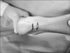
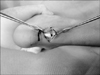
Step 1. The first step is to identify two to three deep creases of the skin about 2 cm apart over the Achilles tendon. When there is a mild to moderate equinus deformity, one skin crease of 2 to 3 cm above the tendon insertion is chosen (Fig. 1). When the contracture is severe (more than 20°), one or two more incisions on the proximal creases are needed for the adequate lengthening.
Step 2. The skin and fascia are carefully incised with the number 15 blade avoiding an inadvertent transverse incision underneath the Achilles tendon. The paratenon is also incised carefully along the skin incision line.
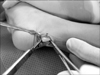
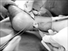
Step 3. The flares are used to separate the paratenon from the Achilles tendon, distally and proximally. The plantaris tendon is identified on the medial border of the Achilles tendon. With the small retractors placed under the paratenon, a distal longitudinal cut is made, beginning in the middle of the tendon. The ankle is plantally flexed to provide the redundant skin. The distal longitudinal tendon incision must be done carefully, in order to avoid laceration of the distal part of the transverse skin incision (Fig. 2). When the tip of the blade touches the calcaneus, the blade is slowly turned 90° medially and the medial 1/2 of the tendon is incised (or a lateral 1/2 incision could be used when necessary, such as in the planovalgus with short Achilles tendon). The plantaris tendon insertion was also cut. Extra cautions must be taken to prevent damage to the tibialis posterior nerve and vessel and to the flexor hallucis longus tendon, which passes near the medial malleolus. The excessive advancement of the blade medially would increase the risk of damage to the neurovascular bundle.
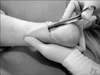
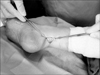
Step 4. For the proximal incision, the surgical scissors are used for the longitudinal cut. A small retractor is placed under the deep fascia and paratenon, and the ankle is dorsiflexed to advance the cut more proximally and to provide tension in the tendon, so the surgeon could feel when the tendon is completely cut. After 3 cm to 4 cm of a longitudinal cut proximally for mild to moderate equinus in children or 5 cm to 6 cm for moderate equinus in adolescents, the scissors are slowly turned laterally and the lateral 1/2 of the tendon is cut. A sudden jerk can be felt when the cut is complete. Extra caution is needed in order to prevent damaging the sural nerve. A thumb (Fig. 3) or a middle finger tip (Fig. 4) was placed over the tip of the surgical scissors to prevent them from advancing beyond the lateral (to protect the sural nerve) or medial border of the tendon. If unable to perform a dorsiflexion of the ankle after the proximal lateral cut, the plantaris tendon needs to be rechecked because it may not have been cut when the distal medial incision was done. When deformity is severe, the second and the third proximal longitudinal incisions of the tendon are made to create a longer lateral strip.
Step 5. The amount of lengthening required is determined with the medial and lateral strips of the tendon, held side by side with the thumb forceps while applying moderate tension after the ankle has been maximally dorsiflexed once or twice (Fig. 5). With a surgical pen, the level of the distal end of the medial strip, which is displaced proximally following the maximal ankle dorsiflexion, is marked on the lateral strip with the ankle in the neutral position (Fig. 6). A simple 2 layer, anterior and posterior suture technique is used. The first is from the anterior side of the tendon, starting the suture and burying the knot inside the tendon (Fig. 7). The same technique is applied on the posterior side (Fig. 8). The consistent suturing is important to prevent a palpable lump on the skin after the surgery, especially on the posterior aspect of the tendon.
Step 6. For the accurate suturing of the paratenon and fascia, it is important to identify them clearly. The first stitch has to be done carefully to avoid suturing these two structures together with the Achilles tendon located below them. The suture binds the paratenon and the deep fascia together, and then a subcutaneous skin suture is applied.
After Treatment
A short leg cast with the ankle at 90° is applied for 6 weeks in the patients under 10 years of age. For additional protection, either a second short leg cast or an ankle foot orthosis is applied for 4 to 6 weeks in the patients over 10 years of age. The gentle active and passive ankle dorsiflexion and heel lifting exercises are gradually recommended after immobilization. Any participation in active sports are permitted after another 6 weeks.
Assessment of Results
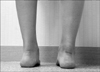
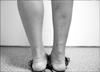
We evaluated the dorsiflexion angle of the ankle before the surgery and at the latest follow-up. The time under anesthesia for the lengthening procedure and any of the postoperative complications were also noted. The surgical results were evaluated using the American Orthopaedic Foot & Ankle Society (AOFAS) Ankle-Hindfoot scale. In addition, to evaluate the patients' satisfaction regarding the surgical scar, we allotted another 10 points to the cosmesis of surgical scar (Fig. 9). For this component, we only have the score after the surgery.
RESULTS
Results of Z-lengthening of the Achilles Tendon with a Transverse Skin Incision
The average improvement in dorsiflexion was 26.9° (range, 15° to 38°). The mean time under the anesthesia was 30.6 minutes (range, 20 to 40 minutes); the actual duration of the surgery was approximately 15-20 minutes. All of the patients showed improvement in gait pattern and they showed satisfactory cosmetic results. The mean AOFAS score improved from 56.1 to 81.8. Within this group of patients, those with neuromuscular or other disorders showed low AOFAS scores, which improved from 42.3 to 67.1, compared to those with idiopathic Achilles tendon tightness, which improved from 69.9 to 96.5. As for the patients' satisfaction with the transverse skin incisions, the mean score was 7.2 points out of 10 points (Fig. 10). No patients had complications such as the tendon adhesion, total transection, excessive lengthening (calcaneus deformity), or neurovascular damage. The recurrence of the tendon shortening was noticed in 4 ankles of the 2 cerebral palsy patients, and the second lengthening procedures were performed through the same incisions. The pain in the tendon suture site was observed in one foot.
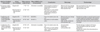
Comparison with Percutaneous Sliding Lengthening and Z-lengthening with a Medial Longitudinal Incision
The recurrence and other complications were noted for all 3 techniques (Table 1). Our new technique attempts to take the best points from both the percutaneous sliding lengthening and Z-lengthening (with a medial longitudinal incision) techniques.
DISCUSSION
The Achillles tendon has three sources of blood: the posterior tibial artery in the distal section, the peroneal artery on the mid-section, and the posterior tibial artery in the proximal section.15,17,18) The arteries of the Achilles tendon and its paratenon are oriented in three directions: longitudinal, transverse, and deep. In most cases, the larger arterial branches run on the surface of the tendon transversely, perpendicular to the direction of the tendon fibers. Our transverse distal incision is made on a natural skin crease, with its length from 1.0 cm to 2.0 cm depending on the size of the patient. Compared to the longitudinal incision, this minimizes the risk of damaging the arterial vessels running transversely on the posterior surface.
The anatomical structures at the risk of damage, with our technique, are the posterior tibial nerve, the artery located in the posteromedial area of the ankle, and the sural nerve located laterally from the Achilles tendon. At the level of the ankle joint in an adult, the posterior tibial nerve is located at an average of 11.8 ± 2.4 mm and the posterior tibial artery at 16.7 ± 3.8 mm, both anterior from the Achilles tendon.19) These structures are, on average, less than 1 cm from the nearest margin of a given percutaneous triple-hemisection in the adult.11) The sural nerve crosses the lateral border of the tendon at an average of 9.8 cm (range, 7 to 16 cm) from the calcaneus in the adult.20,21,22)
All of these distances are shorter in children. The two useful tips to avoid damaging these structures are as follows. First, a small retractor is placed at the medial side of the tendon when the blade is turning 90° to cut the medial 1/2 of the tendon at the distal insertion site. Second, the thumb or the middle finger tip, depending on which hand the physician uses to support the patient's lower leg, is placed at the lateral margin of the tendon when the proximal lateral cut is performed. This will prevent the further advancement of the surgical scissors' tip toward the lateral side, so the damage of the lateral sural nerve can be avoided.
It is difficult to quantify the amount of corrections required.7,23) With our technique, preservation of the peritendinous attachments in the proximal tendon prevents the excessive proximal migration of the medial or lateral strips of the tendon, even with the maximal dorsiflexion of the ankle after Z-lengthening, because only the posterior aspect of the tendon is exposed transversely. The foot is maximally dorsiflexed several times and then positioned in neutral, while the tendon is sutured under a moderate tension obtained by thumb forceps on both halves of the tendon. The toe flexors are carefully examined at the same time as the maximal ankle dorsiflexion to see whether they are also contracted. The flexor lengthening was performed when it was indicated, such as the flexor tenotomy at the proximal interphalageal joint in muscular dystrophy and trauma patients, and the tendon recession in the toe flexor and tibialis posterior at the level of the musculotendonous junction in cerebral palsy patients.
Our technique can provide the medial and lateral strips of the sufficient lengths with a single incision in the mild to moderate ankle equinus. It is desirable to have more than 1 cm in children or 1.5 cm in adults of overlap of the medial and lateral strips for a firm side by side suture with adequate correction. Even suturing with the knots buried inside the tendon is essential to produce a smooth posterior surface. In a cross-section of the Achilles tendon, the insertion of the tendon is flat and spread-out as it attaches to the tuberosity of the calcaneus, while the midsection is oval shaped and is narrower than its insertion and the origin.15) Therefore, the partial excision of the thickened part of the distal tendon stump to match the proximal side is necessary to avoid a palpable lump after the suture. It is also important to suture the paratenon and the deep fascia together with the knot buried in the soft tissues, using an absorbable suture material. A special care must be taken with the first suture to avoid suturing these structures with the underlying Achilles tendon. Also, the suture should be even without crumpling the skin.
In the case of a severe equinus deformity, we apply a cast with the ankle in a mild equinus position while making sure there is an adequate vascular supply in the skin. One week after the surgery, when the skin circulation is satisfactory, a second cast with the correct ankle position is applied. We also adopt the Ilizarov gradual correction technique to correct a severe ankle equinus deformity. For this, before application of the Ilizarov external fixator, we use 2 or 3 transverse incisions and lengthen the tendon through the second and third incisions without suturing from side to side. The lengthened tendons gradually slide against each other until the deformity is corrected with the Ilizarov apparatus. The short leg cast after 4 weeks of the complete correction and the ankle foot orthosis during the night provide a good result and prevent recurrences, especially for the patients with neuromuscular disorders, club foot, and trauma. The correction happens quickly, without any significant pain or discomfort to the patient as the tendon lengthens. None of our patients, even those with severe deformity, have shown any skin problems due to the tension.
We retrospectively studied the results of our new technique and compared them with the results of two other techniques we have used before. The patient attributes, which include the number of patients, ages, causes, severities, etc., are not the same for the different techniques. Our new technique seems to have a low rate of recurrence of equinus deformity (4 of 95 feet) at the medium-term follow-up, compared to the other approaches; and no patient with a crouch gait resulted from the over-lengthening. We believe that this was due to the short skin incisions, the preservations of the paratenon and the deep fascial tube, and the use of Z-lengthening rather than the open or percutaneous sliding lengthening or lengthening the aponeurotic tendon of the gastrocnemius. With the new approach, we had fewer patients complaining of the pain compared to those who were operated with a longitudinal skin incision. The cases of recurrence were mostly limited to the cerebral palsy patients, who were less than 5 years old at the time of surgery, and they appeared at least 3 years after the surgery. Although the range of the passive dorsiflexion was decreased during the period of rapid growth in these children, any recurrent equinus was easy to treat because there was little scarring at the operation site.
In summary, our technique of Achilles tendon lengthening has proven to be an improvement over the other techniques we have employed, in terms of surgical time, tendon healing, and complication rates. This is perhaps due to the small skin incision and the conservation of the paratenon and the deep fascia.

 XML Download