

 PDF
PDF ePub
ePub Citation
 Print
Print


The wrist is a complex joint consisting of bony structures, including the distal radius, distal ulna, and eight carpal bones and a ligament complex that divides extrinsic and intrinsic ligaments. Therefore, the prognosis of wrist injuries can be affected by soft tissue injuries as well as fractures.1-4) Distal radial fractures (DRFs) are common injuries that occur in the upper limb and are treated using various methods, including closed reduction and cast immobilization, percutaneous K-wire fixation, external fixation, intramedullary fixation, and open reduction and internal fixation. Various clinical results and complication rates have been reported.4,5) However, previous studies have been mostly limited to fractures that occur in the distal radius with associated fractures in the distal ulna. Based on the development of arthroscopic instruments and techniques, DRFs associated with soft tissue injuries such as the triangular fibrocartilage complex, scapholunate ligament, and lunotriquetral ligament have been reported. Some studies have reported that associated soft tissue injuries may affect the outcome of DRFs.1-4)
As soft tissue injuries may be accompanied by DRFs, carpal bone fractures (CBFs) may occur in association with DRFs. Avulsion fractures of carpal bones are commonly observed in patients with DRFs and may indicate injuries to the intrinsic or extrinsic ligaments of the wrist. Additionally, CBFs may be untreated, because CBFs are frequently missed on initial radiographs, which may lead to persistent pain or subsequent wrist dysfunction and eventually affect the outcome of DRF treatment. If associated CBFs are misdiagnosed and left untreated, unsatisfactory DRF clinical outcomes may occur. Therefore, associated carpal injuries must be ruled out for DRFs.6) However, the frequency and distribution of CBFs associated with DRFs have not been reported, since cases of scaphoid fractures accompanying DRFs are usually reported. The purpose of this study was to investigate the frequency and distribution of CBFs associated with DRFs.
METHODS
A total of 313 patients who underwent operative treatment after being diagnosed with a DRF by plain X-ray between March 2007 and January 2010 were reviewed retrospectively. In this study, 223 patients who underwent preoperative computed tomography (CT) were included. DRFs that had a fracture-dislocation associated with a CBF or suspected intercarpal instability on plain X-ray were excluded.
There were 82 males and 141 females with a mean age of 57.6 years (range, 16 to 97 years). The mechanism of injury included fall on an outstretched hand (143 cases), fall from height (38 cases), and traffic accident (42 cases). All DRFs underwent anteroposterior, lateral, and oblique plain X-ray views of the wrist prior to and after reduction. CT scans were performed in neutral forearm rotation after wearing a sugar tongs splint. The CT machine was an Mx8000-IDT instrument (Philips, Eindhoven, The Netherlands). A 1 mm interval sectioned image was obtained and reconstructed into sagittal, axial, and coronal images using the MxView ver. 3.5 program (Moxa, Brea, CA, USA).
All DRFs were investigated for the frequency and location of associated CBFs on CT scans. The DRFs were classified by the arbeitsgemeinschaft für osteosyntheses (AO) classification,7) and the frequency of associated CBFs was checked for each AO classification. The DRFs were divided into two groups: DRFs with CBFs and those without CBFs. Differences in age, sex, and body mass index between the two groups were compared with the chi-square test. The mechanism of injury was divided into a low-energy group including fall on an outstretched hand and a high-energy group including fall from height and traffic accident. The frequency of associated CBFs between the two groups was compared with the t-test. The frequency among AO types was compared with Fisher's exact test.
RESULTS
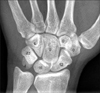
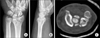
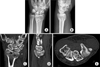
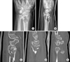
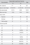
Among 223 DRFs, 46 were accompanied by CBFs. There were 20 males and 26 females with a mean age of 57.3 years (range, 16 to 87 years). The mechanisms of injury included fall on an outstretched hand (30 cases), fall from height (11 cases), and traffic accident (five cases) (Table 1). The distribution of CBFs was 23 cases in the triquetrum, 16 in the lunate, 12 in the scaphoid, five in the hamate, and four in the pisiform (Fig. 1). Among the 46 DRFs with CBFs, fractures of the triquetrum were the most commonly identified, and most CBFs were identified in the proximal carpal row. Also, among the 46 DRFs with CBFs, cases with associated fractures of one carpal bone (Fig. 2) occurred in 36 case, two in seven cases (Fig. 3), three in two cases (Fig. 4), and four in one case (Fig. 5). Ten of the 46 cases were accompanied by CBFs in more than two carpal bones.
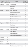
All DRFs were classified by the AO classification, and the frequency of associated CBFs was checked for each AO classification (Table 2). Type A3 was complicated in two of 44 cases, type B1 was complicated in two of 11, type B3 was one of 12, type C1 was one of 13, type C2 was 19 of 75, and type C3 was 21 of 66.
No significant differences were observed for age (p = 0.38), sex (p = 0.30), or body mass index (p = 0.28) between patients with DRFs and CBFs (46 cases) and those without CBFs (177 cases). No significant difference was observed in the frequency of associated CBFs between the low-energy and high-energy groups separated by the mechanism of injury (p = 0.86). Despite the relatively high frequency of associated CBFs in AO types C3 or C2, no difference was found among AO types (p = 0.18).
DISCUSSION
The eight carpal bones composing the wrist joint are of various sizes and shapes, and stability among carpal bones is maintained by extrinsic and intrinsic ligaments. Due to this complex anatomical structure, the diagnosis of CBFs using plain radiographs is difficult. Misdiagnosed or untreated CBFs can lead to complications including nonunion, malunion, avascular necrosis, carpal instability, articular incongruity, or traumatic arthritis and can consequently induce persistent pain and result in functional compromise.8-10) In this study, we confirmed associated CBFs in 20.6% of DRFs that had been surgically treated. However, plain radiographs were insufficient to make a diagnosis, thus most cases were confirmed by CT. Welling et al.10) suggested that CT should be considered if clinically warranted, because 30% of wrist fractures are not detected on plain radiographs. In addition, as other CBFs, except those of the scaphoid, are more difficult and have lower sensitivity for detection on plain radiographs, the need for specific radiographic views or CT is emphasized. Kiuru et al.11) also recommended the need for CT to assess complex wrist fractures or when initial radiographs are equivocal, because only plain radiographs may miss occult fractures, which usually occur in small carpal bones. In this study, only 14 of 46 DRFs associated with CBFs were detected on plain radiographs and all the others were found on CT scans.
The frequency of CBFs of all hand injuries ranges from 8% to 19%.8,9,12,13) The scaphoid accounts for about 70% of all CBFs, and the remaining carpal bones account for about 30%. More than 90% of CBFs are found in the proximal carpal row. In our study, 89% of CBFs associated with DRFs occurred in the proximal carpal row. However, the frequency of fractures of the triquetrum and lunate was higher than that of the scaphoid, and only the hamate was injured in the distal carpal row. Accordingly, the frequency and distribution of CBFs associated with DRFs were different from those of ordinary CBF cases.
DRF is one of the most common fractures in the upper extremities which results from a fall on an outstretched hand with an extended wrist. Because the mechanism of carpal bone injuries is similar to that of DRFs, CBFs may often occur simultaneously with DRFs. The complication rates of DRFs in previous studies vary from 6% to 80%.4) The clinical result of DRFs may be influenced by injury severity, any associated soft-tissue trauma, and treatment method. In accordance with the development of arthroscopic procedures for the wrist joint, associated damage to soft tissues such as the triangular fibrocartilage complex or intercarpal ligaments has been reported, and the importance of treating this accompanying damage has been emphasized.1-3) However, the prevalence and effect on clinical results of CBFs associated with DRFs have not been reported. Most reports regarding simultaneous CBFs and DRFs are scaphoid fractures, which account for 0.75%-6.5% of all DRFs. Stable internal fixation of scaphoid fractures and DRFs is generally performed for early mobilization of the wrist joint and to avoid complications such as scaphoid nonunion.14,15) Pretell-Mazzini and Carrigan6) reported simultaneous DRFs and carpal bone injuries in children. They emphasized that orthopedic surgeons must first rule out CBFs because the mechanism of injury is similar for both fractures, and inappropriate treatment may lead to unsatisfactory outcome. Most other CBFs, except scaphoid fractures, can be treated by immobilizing the patient for 4-6 weeks. In our study, all CBFs except five waist fractures of the scaphoid were conservatively managed. In accordance with the development of a locking plate for DRFs, however, early mobilization of the wrist joint is often performed after the operation for the DRF.16,17) If a CBF is not found on the initial plain radiograph and early exercise is performed after stable internal fixation of the DRF, the undetected CBFs may affect the patient's rehabilitation and treatment outcome. We generally immobilize all DRFs for about 4 weeks after surgery using a locking plate. In a comparison study between an early motion group and a late motion group by Lozano-Calderon et al.,18) no significant differences in range of motion, grip power, or clinical scoring were observed. Therefore, we believe that immobilization for an appropriate period after surgery is necessary if associated CBFs are detected or uncertain on the initial plain radiograph. Orthopedic surgeons must carefully access associated CBFs on the initial radiography or CT, which are observed as avulsion fractures and may imply ligament damage or instability of the wrist joint.4,19-21)
The strength of the present study was that it investigated the frequency and distribution of CBFs associated with DRFs, which have not been reported previously. However, several limitations should be noted. First, there was a possibility of selecting more severe fractures, because surgically treated DRFs were included and those without CT were excluded. Accordingly, the frequency of CBFs may be higher than observed due to selection bias. DRFs that are conservatively treated should be included in the materials of the study to overcome this bias. However, it should be noted that diagnosing CBFs on plain radiographs is difficult. Second, DRFs with associated CBFs were surgically treated with various methods such as volar plate fixation (29 DRFs), dorsal plate fixation (five DRFs), external fixation (seven DRFs), and percutaneous K-wire fixation (five DRFs). Because these fractures were not treated by one surgical method with the same postoperative protocol, they did not have a significance to evaluate the clinical outcomes. Third, radiological parameters of the distal radius were not evaluated, as the main purpose of this study was to investigate CBFs complicated by DRFs. Forth, we did not compare clinical outcomes between patients with DRFs and CBFs and those without CBFs. A further study is required to evaluate whether associated CBFs in DRFs affect the clinical outcome of DRFs.
In conclusion, DRFs were accompanied by CBFs at a considerably high frequency, and most of the fractures were combined in the proximal carpal row. The frequency of CBFs increased in severe fracture types such as AO types C2 and C3, and 90% of the DRFs with associated CBFs were classified as these two types. If DRFs with associated CBFs are conservatively treated, no additional attention to the CBF, except the scaphoid, may be required. In contrast, if a DRF is surgically treated and early mobilization is planned, misdiagnosed or untreated associated CBFs may affect the final outcome. Therefore, confirmation of a CBF accompanied by a DRF is required along with use of CT scan for the detection of CBFs complicated by DRFs that require operative management, as plain radiographs are insufficient for a definitive diagnosis of associated CBFs.

 XML Download