

 PDF
PDF ePub
ePub Citation
 Print
Print


Recurrent extensor carpi ulnaris (ECU) subluxation is one of the disorders that provoke ulnar wrist pain, which has been rarely reported. It normally occurs due to sports activities, such as golf or tennis. The characteristic symptom is a snapping sound with ulnar wrist pain particularly in rotation of the forearm. The treatment can be conservative but sometimes it requires surgical treatment. We encountered a case of ECU dislocation combined with extensor tendon subluxation of the long finger at the metacarpophalangeal (MP) joint. To our knowledge, there has been no report of simultaneous ECU dislocation with extensor tendon subluxation. Therefore, we report these results with a review of the relevant literature.
CASE REPORT
A twenty-year-old male patient presented to our clinic with pain of the right wrist and the long finger MP joint. Two years earlier, the patient slipped on his hand. After injury, he did not received any evaluation or treatment, but he felt a snapping of the ulnar side of the wrist in forearm rotation and dorsal side of the long finger MP joint in MP flexion. At that time, the symptoms improved spontaneously after several weeks without specific treatment, three months earlier, after heavy lifting, he had persistent symptoms despite the conservative treatment including short arm splint for 4 weeks. A physical examination revealed painful ECU dislocation in forearm rotation and painful extensor tendon subluxation at the long finger MP joint in MP flexion. He showed generalized ligament laxity according to the Beighton and Horan criteria but did not have any symptoms in the contralateral hand.1)
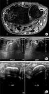
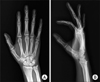
Plane radiography did not reveal any fracture or abnormality. Ultrasonography and magnetic resonance imaging (MRI) were performed to confirm the subluxation and identification of the associated pathology. In dynamic ultrasonography, the sagittal band (SB) was subluxated ulnarly as the long finger MP joint was flexed more than 70°. In addition, the ECU was subluxated volarly in forearm supination with tendon attrition at the level of the ulnar styloid process where the ECU groove exists. MRI revealed longitudinal splitting approximately 5 cm at the level of the ulnocarpal joint and dislocation of volar half slip of the ECU tendon on T2 axial plane image (Fig. 1).
The patient had been in military service. He required surgical treatment for rapid return to his previous work. Therefore, surgical treatment was performed.
With the patient under general anesthesia, the long finger MP dorsum was approached longitudinally. The radial SB did not reveal any definitive rupture but diffuse thinning was observed. The extensor tendon slipped off the metacarpal head ulnarly as the joint flexed intraoperatively. The redundant radial SB was augmented using a cross-stitch with a 6-0 prolene suture (Fig. 2A). This effectively prevented any additional slippage of the extensor tendon of the long finger.
With an approximately 4 cm longitudinal incision in the ulnar styloid area, the ECU sheath on the ECU groove was approached protecting the dorsal ulnar nerve sensory branch. Neither the ECU sheath nor extensor retinaculum was ruptured but the space within the ECU tendon compartment was dilated because of its redundancy at the ulnar styloid process. An ECU tendon dislocation in an ulnar and palmar direction was confirmed with the forearm in supination and the wrist in palmar flexion. The fibro-osseous sheath of the ECU groove was incised at the ulnar border and elevated radially. The ECU tendon surface showed no longitudinal tear unlike the MRI finding. The ECU groove was deepened approximately 2 mm using a curette. Two small FASTak (Arthrex Inc., Naple, FL, USA) suture anchors were inserted at the ulnar border of the ECU groove just proximal to ulnar styloid and 1 cm apart proximally. The overlying ECU sheath was advanced to the underlying periosteum reducing the enlarged sheath volume with a horizontal mattress tie (Fig. 2B). A long arm splint with MP extension at neutral forearm rotation was applied for 4 weeks and the wrist motion was allowed thereafter. A removable splint was applied for an additional 2 weeks. Vigorous activities were restricted for an additional three months. At the 1-year follow-up, the long finger MP joint, wrist and forearm showed normal motion with no pain (Fig. 3). He returned to his previous activities without a recurrent dislocation of the ECU tendon and long finger extensor tendon.
DISCUSSION
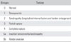
The ECU tendon is encased by a fibro-osseous sheath deep to the extensor retinaculum as described by Spinner and Kaplan, which contributes to distal radioulnar stability at the distal ulnar groove. A classification of the different possible lesions of the ECU tendon and osteofibrous sheath was reported on previously published series.2,3) In this case, the ECU sheath is stripped from the periosteum but remains in continuity, forming a false pouch, and the ECU tendon itself has tendinopathy and several longitudinal splits (Tables 1 and 2).
There are many different conditions triggering ulnar side pain. Classically described maneuver for the evaluation of ECU tendonitis has some disadvantages to identify ECU problem. ECU synergy test helps to identify chronic dorsal ulna-sided wrist pain and has high diagnostic accuracy. In our case, pain along the dorsal ulnar side did not provocated by this test.
Imaging studies, such as MRI for the diagnosis of the ECU subluxation are nonspecific except for the tenosynovitis but dynamic ultrasonography is useful for identifying the ECU dislocation in the forearm rotation with ulnar deviation and palmar flexion.4) In this case, dynamic ultrasonography also showed a signal void about 70% at the distal ulnar ECU groove. In addition, MRI showed longitudinal splitting with a dislocation of the volar half of the ECU tendon, which suggests static dislocation of the volar half of the ECU tendon in neutral forearm rotation. Therefore, a decrease in the ECU tendon volume with a longitudinal tear within the ECU groove might be a diagnostic clue of ECU volar subluxation (Fig. 1).
The timing and appropriate treatment for the recurrent symptomatic ECU subluxation is controversial. Recently, this incidence of injury has increased, particularly in athletes and the active young population. Therefore, more active treatment might be needed in these patients.
Surgical treatments include direct repair and reconstruction of the ECU sheath with extensor retinaculum flap or fascial patch. The anatomic ECU tendon sheath reconstruction technique using suture anchor has shown good outcomes in 21 patients.5)
This case had concomitant extensor tendon subluxation at the long finger MP joint. The patient had symptoms in the absence of any definitive tear of ECU sheath or SB. Therefore, patients with generalized ligament laxity, as in this case, should be treated carefully and warned at the initial trauma event of recurrent dislocation. Surgical stabilization should be considered, if the symptoms recur despite conservative treatment.
When the wrist joint moved to ulnar deviation and palmar flexion with active contraction of the ECU, the ECU tendon has translational stress to be forced out of the groove to the volar-ulnar side in terms of the ECU muscle force vector traversing the wrist joint. The ulnar styloid process serves as a pulley around which the ECU tendon changes direction to transmit the force to the 5th metacarpal base.
An ultrasound imaging study of the ECU tendons of 40 symptom-free wrists of healthy volunteers showed that the mean ECU tendon displacement was greatest in the supinated forearm position followed by the neutral position and was least in the pronated position to a mean of 1.46 mm (SD, 2.03) beyond the apex of the ulnar border of the ulnar groove.6)
On the other hand, the dorsal edge of the ECU tendon was never dislocated over the ulnar border of the groove despite asymptomatic physiologic subluxation. The forearm rotation is believed to change the gliding resistance of the ECU tendon or potential compartment volume within the ECU compartment. For the extensor digiti minimi tendon, the gliding resistance in pronation was higher than that in neutral or supination.7) Therefore, the forearm supination might increase the potential volume of the ECU compartment, resulting in a decrease in gliding resistance of the ECU tendon. If the ECU sheath is attenuated, the ECU tendon does not maintain its normal position.
The other possible explanation for the ECU subluxation is the ulnar variance depending on forearm rotation. If the ulnar styloid, as a pulley for the ECU tendon, is shortened relative to the distal radius, the ECU tendon distal to the ulnar styloid might have subluxation tendency volar to the ulnar groove because soft tissue pulley is not as strong as bony pulley.
Acute closed SB injury might be treated conservatively, such as digital splinting, but surgical treatment might be necessary in a chronic SB injury. Many surgical techniques have been described depending on the SB deficiency.8,9) In this case, the radial SB has a laxity without a definitive rupture. We consider that repetitive use causes chronic pain and it is not related with initial trauma. Therefore, neither additional reinforcement nor reconstruction is needed except for a cross-stitch suture only for redundant radial SB.
The ultrasound findings of a ruptured SB showed focal hypoechoic thickening of the SB at the side of the extensor tendons, even though the normal thickness of the SB has a wide range of values.10) The disappearance of the normal architecture of the SB as well as the extensor tendon subluxation to the contralateral side is also noticed.11)
Patients with generalized ligamentous laxity might experience suboptimal results of the ligament stabilizing procedure because of the inherent connective tissue extensibility as well as laxity of the secondary restraints caused by the composition of the connective tissue and the orientation of the various soft-tissue structures. On the other hand, in this case, the patient's symptoms were relieved by a retinaculum augmentation procedure using his own tissue. Therefore, retinaculum augmentation should be considered primarily for recurrent ECU subluxation and extensor tendon subluxation, even though the patients show generalized ligamentous laxity.


 XML Download