

 PDF
PDF ePub
ePub Citation
 Print
Print


In Pakistan, musculoskeletal oncology is yet to develop as an individual orthopedic subspeciality. Most bone sarcoma patients are treated either with an amputation or with a general orthopedic surgeon, whose practice experience varies regarding the management of tumor, its resection and reconstruction. Hence the oncological outcomes cannot be viewed as satisfactory. As in most other developing countries, patient-presentation to medical facilities is often delayed for various reasons. Presentation with a huge mass with or without metastasis is very common. Possibility of performing a limb salvage surgery poses significant surgical risks even in the hands of subspecialised orthopedic surgeons. Most bone sarcomas are typically metaphyseal, and the delayed presentation brings tumors closer to the joint line. A resection of the joint and arthrodesis leaves the patient with a significant functional disability, as tumor prosthesis is not affordable for most of the patients. Our efforts are thus focused on saving the patients' respective joints and providing best possible functional outcome after a biological reconstruction of the skeletal defect. This has entailed us to err on the side of a narrow margin of resection towards the side of the joint, relying heavily on the response of neoadjuvant and adjuvant chemotherapy.1,2)
We have performed the surgeries only in cases where the tumor has not crossed the epiphyseal scar. After resection, we are usually left with a small area of metaphyseal bone distal to the epiphyseal plate. We have found locking compression plate (LCP) very useful in such situations. Its ability to offer a fixed angle construct in the locking-hole reduces the toggle between the plate and screw and theoretically decreases the incidence of implant-related failures.3) This would seem to be advantageous in autoclaved (our preferred choice) or allograft bones, because time-to-healing is prolonged in such avascular bones as compared to normal fracture healing.4,5) Rigid fixation with the option of unicortical screw purchase in the remaining skeletal tissue could help reduce stress risers and subsequently, the risk of fracture, another mode of failure in both autoclaved and allogeneic bones. All of this also holds true in the fixation of pathological fractures, where the whole bone is often quite weak and studded with lytic lesions from the metastasis present all across its length.
In this study, we have evaluated the role of LCP in the reconstruction of bone following a tumour resection, as well as in fixation following pathologic fracture.
METHODS
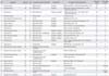
This is a retrospective analysis of patients with bone tumors, benign as well as malignant, in whom surgical resection or fixation of pathologic fracture was done using a LCP. Twenty five patients were operated upon during the period of January 2008-January 2010. Curative surgery was done in 17 patients in whom limb salvage procedure was performed. Reconstruction was done using fibular graft, synthetic beta tri-calcium phosphate granules and/or autoclaved bone, with the skeletal stabilization done using a locked compression plate. Internal fixation of pathologic fractures was done in the remaining eight patients. Patients with a completed follow-up of at least one year have been assessed for the evidence of union at resected ends and pathologic fracture sites. Both early and late complications were noted in all patients. Details of the patients are summarized in Table 1.
RESULTS
A minimum follow-up of 1 year was available in all patients, and they were subjected to the assessment of union. Out of twenty-five, seventeen are female and eight male. Nineteen patients had a reconstruction done for malignant pathology; and remaining six patients included four with a giant cell tumour and one each with aneurysmal bone cyst and fibrous dysplasia. Mean age at the time of surgery was 30 years (range, 7 to 72 years). Mean time for union was 6.5 months, achieved in 17 patients at an average of 6 months after reconstructive surgery and 4.75 months after fixation of pathological fractures.
Local complications were noticed in three patients. One patient had an early wound infection which required debridement without removing the plate, another developed a nonunion and a third had a periprosthetic fracture. Systemic complications were also present, which included acute renal failure and postoperative myocardial infraction in two patients, with both requiring a prolonged length of hospital stay. Both patients are doing well now.
DISCUSSION
Limb-sparing resection and reconstruction have become the treatment of choice in extremity malignancies. If done with adequate margins and minimum surgical morbidity, along with neo- and adjuvant chemotherapy, results of limb-sparing surgeries are no worse than amputation, both in terms of recurrence and spread of disease. Limb preservation is also functionally and cosmetically superior to amputation. A successful treatment requires the combination of surgical eradication and each patient's specific functional and aesthetic rehabilitation.6,7)
Reconstruction after tumor excision is a challenge, especially in skeletally immature patients. Skeletal reconstruction faces difficulties due to mechanical factors such as short residual periarticular proximal or distal segment and the proposed use of bone cement and arthrodesis after treatment of the skeletal defect. Biological obstacles include poor quality of bone due to tumor and secondary effects of radiotherapy or chemotherapy.8,9)
Reconstructive options after resection of bone tumors around knee-joint include endoprosthesis, arthrodesis with long intramedullary nail and conventional dynamic compression plate. Tumor prosthesis is one of the most common and successful solutions for reconstruction following a resection of bone tumor located to the metaphysis of long bones. A large exposure of tissue planes during this type of surgery, dissection across vascular distributions, malnutrition and immune-compromised conditions of the patients all contribute to the high risks of wound dehiscence and subsequent infection following endoprosthetic reconstruction.10) Financial constraints on the part of our low-income patients limit the use of endoprosthesis as a routine for such reconstructions.
Traditionally locking plates have been used in the fixation of pathological fractures and allograft fixation after tumor resection.9) We have used locking plates in skeletal reconstructions. There are very few reports in the literature regarding the use of LCP in orthopedic oncology. These locking plates are fixed-angle devices with high pull-out screw strength, load sharing and elastic properties, which make them superior to the traditional counterparts.11,12) These plates make for an easier reconstruction and allow better fixation in poor-quality bones, especially weakened by metastasis at multiple sites and pathologic fractures and/or following a chemo- or radiotherapy.13)
We used the titanium locking plates which are compatible with the required follow-up magnetic resonance imagings (MRIs). This should make for an early and easy detection of tumor recurrence on follow-up MRI scans, which was not possible in the previous stainless steel plates. The minor disadvantages include the requirement for a long length of plate, difficulties in contouring and implant prominence in limb salvage surgeries. We did not encounter any of these problems in our patients.
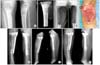
Stabilization of pathologic fractures and autoclaved bone (or allograft) fixation after resection of an osseous tumor are two areas where a strong internal fixation is critical to successful healing. Pathologic bone presents many challenges to the surgeon, as the quality of the bone is compromised from a destructive oncologic process, irradiation or chemotherapy.8) This creates significant biological barriers for cellular response and tissue repair, and can prolong the time to union. As long as the patient's life expectancy and overall health are adequate, a surgical intervention is often necessary to treat these fractures.14) Due to a limitation of resources, we use autoclaved tumor bone in most of our biologic reconstructions following tumor resections.15) Such autoclaved bone (or allograft) reconstructions also present a challenge to osteosynthesis. As these do not have a native blood supply, they almost always demonstrate a delayed time to union. The biologic inertness of autoclaved or allograft bone combined with attenuated host bone secondary to malignancy, periosteal stripping, chemotherapy or irradiation creates a challenging environment for healing. Mechanical obstacles, including long or short segments, multiple implants and proximity to cement, also negatively impact bone healing and place unique demands on the chosen implant for fixation. Locking plates effectively address the problem of short bone segments by providing a substantial amount of stability over a small surface area by giving multiple fixed angle points for fixation (Fig. 1).7)
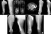
Complications such as periprosthetic fracture, breakage of plate, prominent implant, non-union at host-graft junction and bone and joint stiffness have all been reported in literature. In our series, we have encountered one patient with non-union who had undergone a limb salvage surgery of distal femoral osteosarcoma with a wide margin excision and reconstruction with combination of nonvascularized fibula with synthetic graft (Fig. 2). In addition polytetrafluroethylene (PTFE) graft was also used to repair femoral artery as a part of the superficial femoral artery was encased in the tumor and thus had to be resected. Non-union was established 10 months post surgery. Femur was subsequently stabilized with a longer LCP and bone grafting, and subsequent healing was evident in 4 months.
Another patient with metastatic cancer of breast presented with pathological fracture of humerus. She had the initial stabilization done with LCP four months prior. She then presented in the outpatient clinic with swelling, pain and gross motion at the fracture sight. X-rays confirmed a fracture just above the plate. The patient was managed with a longer LCP fixation and eventually healed 5 months after the second procedure.
A comparative study in pediatric population showed union in 13.1 month in 75% of the patients fixed with a locking plate, compared to 14.6 months with the standard compression plates. Additionally three patients developed nonunion in the former group, out of whom two healed after autogenous bone grafting, with the third patient requiring a revision of plate for persistent nonunion.16) Another study from India reported the union time of one-month following fixation of pathologic fracture and 120 days after limb salvage surgery.17) In our study, the average union time for curative resection and reconstruction and osteosynthesis in pathologic fracture was 6.5 and 4.75 months, respectively.
The main limitation of this study is its limited sample size. The results are presented mostly as an audit with a limited control over the confounders.
The use of locking plates provided stable fixation that expedited union and allowed for early mobilization of joints above and below, in all our patients. This early healing also allowed for early weight-bearing. Studies with a long-term follow-up and larger series are needed to further assess the utility of these implants. Our early results clearly show the benefits of locking plates with respect to the ease of surgical technique, and the fixation of compromised bone shows this to be a viable and attractive option in the field of orthopedic oncology.
 XML Download