

 PDF
PDF ePub
ePub Citation
 Print
Print


Acromioclavicular (AC) joint injuries are one of the most common shoulder injuries seen in orthopaedics, especially among athletes participating in contact and collision sports. Rockwood et al.1) has classified these injuries into six types, based on increasing severity of injury sustained by the AC ligaments, coracoclavicular (CC) ligaments, and supporting musculature (deltoid and trapezius muscles). Types I and II injuries maintain joint alignment and are successfully treated non-operatively, with the majority of patients able to return to sport at pre-injury level. While type III injuries remain controversial, surgical intervention is recommended for types IV-VI.
Several different surgical techniques have been described in the literature to address AC joint pathology, including primary repair of the CC ligaments, distal clavicle excision, stabilization with metallic hardware, as well as augmentation with autogenous coracoacromial (CA) ligament (modified Weaver-Dunn procedure), allograft, or suture material.2-14) Unfortunately, many of the current techniques have been associated with complications, including residual instability with loss of reduction, failure of hardware, bony erosion, and continued pain.13) One potential reason for failure is that the majority of techniques solely addresses the CC ligaments, without concomitant reconstruction of the AC ligaments, despite biomechanical studies that have shown the importance of the AC ligaments in restraining anteroposterior translation.15-19) Few studies have reported techniques on reconstructing both the CC and AC ligaments.20,21)
We have previously reported a new technique to reconstruct the AC ligament, using an intramedullary free-tissue graft.22) This procedure, in combination with reconstruction of the CC ligaments, using allograft tendons, has shown to be superior to the modified Weaver-Dunn procedure in biomechanical testing.23) However, the Weaver-Dunn procedure does not address the AC ligaments. The purpose of our study was to compare the biomechanical properties of two free-tissue graft techniques that reconstruct both the AC and CC ligaments in cadaveric shoulders, one with an extramedullary AC reconstruction and the other with an intramedullary AC reconstruction. We hypothesized that an intramedullary AC reconstruction would provide greater translational stability and improved load to failure characteristics than the extramedullary technique.
METHODS
Six pairs of matched fresh-frozen human cadaveric shoulders with an average age of 63.8 years (range, 57 to 74 years) were used in this study. The specimens were all from male donors. Each shoulder demonstrated an intact AC joint complex, with no evidence of gross deformity. Each specimen was thawed overnight, the day before testing and then disarticulated at the glenohumeral joint. All soft tissues were dissected free, leaving only the AC joint capsule, CC ligaments, and CA ligament intact.
The scapula was then potted in a rectangular aluminum box, from the inferior border to the level of the glenoid fossa, with the AC joint parallel to the lateral border of the box. The specimen was initially held with two transfixing pins and then augmented with plaster of Paris. The clavicle was potted in 3.2 cm polyvinylchloride piping, medial to the CC ligaments. The clavicle was initially fixed with four unicortical screws and then augmented with plaster of Paris. During all phases of preparation and testing, the specimens were sprayed with normal saline mist, to prevent desiccation.
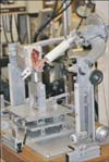
The potted specimens were placed in a custom shoulder testing apparatus that allowed six degrees of freedom, as previously described (Fig. 1).23-26) The scapular box was mounted onto a bearing and lever arm system, with the lateral border face down, to allow compression across the AC joint. Beneath this system were two translational plates, which allowed anteroposterior and superoinferior translation of the acromion with respect to the clavicle joint. During testing, both plates were left unlocked, allowing simultaneous motion in both axes of translation. The potted clavicle was mounted onto the top arc of the apparatus, to maintain anatomical alignment of the AC joint.
AC motion was measured in the anteroposterior and superoinferior directions, using a three-dimensional digitizing system, the MicroScribe 3DLX (Revware Inc., Raleigh, NC, USA). The accuracy and resolution of this device has been determined to be 0.30 mm and 0.13 mm, respectively.26) A reference system was established using the bi-directional translation plates such that X was defined as the anteroposterior direction and Y was defined as the superoinferior direction. During translational testing, the acromion moved relative to the fixed clavicle when a translational load was applied to the scapula. Translational measurements were calculated as the difference between the neutral position of the scapula with no translational load applied and the position of the acromion after a translational load was applied in either the anteroposterior or superoinferior direction. The total anteroposterior and superoinferior translation was then calculated by adding the amount of translation in both directions. The specimens were preconditioned with a 10 N load in the anterior, posterior, superior, and inferior directions for 10 cycles. Translational testing was then performed under six different conditions using a 10 or 15 N load in each direction with a compressive load of 10, 20, or 30 N across the AC joint. Each translation was executed twice to ensure accuracy and the average of the two values was recorded.
The CC and AC ligaments were then sharply transected, while the specimen remained on the testing apparatus. Great care was taken to leave the CA arch intact. One of the specimens was then randomly assigned the intramedullary graft reconstruction while its matched pair received an extramedullary graft reconstruction as described by Grutter and Petersen,20) using semitendinosis tendons. After the reconstruction, the specimens were preconditioned and translationally tested as described above with the native AC joint complex.
The intramedullary graft reconstruction surgical technique previously described was used.22,23,25) Briefly, a 5 cm semitendinosis allograft was doubled over a looped No. 5 Ethibond (Ethicon, Somerville, NJ, USA) passing stitch, while the free ends of the graft were sutured together with Fibertape (Arthrex Inc., Naples, FL, USA) in a Krackow fashion. The end of the Krackow stitch that was oblique to the looped end of the Ethibond was intentionally left long to allow for eventual Fibertape passage through bone tunnels. To create a CC sling, 15 cm of semitendinosis allograft was prepared by placing whipstitches on either end. Using the graft preparation station, both grafts were tensioned to 10 N to remove any subsequent creep.
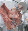
The intra-articular ends of both the clavicle and acromion were opened using sequentially larger drill bits until the intramedullary canal was exposed and able to accommodate 1.25 cm of the graft in either end. Next, 1.5 mm drill holes 1 cm apart were placed into the ends of the blind tunnels at the most anterior and posterior aspects of the superior surface of the distal clavicle and the acromion. The CC sling allograft was passed around the base of the coracoid with the lateral limb anterior to the clavicle and the medial limb posterior to the clavicle. Prior to securing the CC sling, the AC graft was passed through the bone tunnels. Using a Hewson suture passer (Smith and Nephew, Memphis, TN, USA), the looped end of the passing Ethibond stitch was shuttled through the anterior acromion drill hole while the tail end was shuttled through the posterior acromion hole. In a similar fashion, the short tail of the Krackow stitch was passed through the anterior clavicle drill hole, while the long tail was passed through the posterior clavicle drill hole. This long end was then placed through the obliquely located loop of the Ethibond passing stitch and pulled through the acromion. The two ends of the Fibertape Krackow stitch were tied in a cruciate pattern over the reduced AC joint creating an intramedullary graft reconstruction. Finally, the two limbs of the CC sling were tied to each other with three figure of eight stitches using #2 Fiberwire (Arthrex Inc.) incorporating superior clavicle periosteum (Fig. 2).
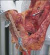
The extramedullary graft reconstruction described by Grutter and Petersen20) in 2005 was used. Approximately 20 cm of semitendinosis allograft with a 4.5 mm diameter was secured with one whipstitch using #2 Fiberwire on one end and tensioned to 10 N to remove any subsequent creep on a graft preparation station. Three 4.5 mm drill holes were made in the clavicle and coracoid to recreate the conoid and trapezoid ligaments. The first hole was drilled in the clavicle from superior to inferior at the junction of the lateral third and medial two-thirds 2 mm from the posterior border of the clavicle. In a similar fashion, the second hole was drilled 15 to 20 mm lateral to the first hole in the midline of the clavicle. The third hole was drilled across the coracoid from anterolateral to posteromedial. Next, two 2.5 mm drill holes were made from lateral to medial in the acromion exiting near the midline of the clavicle. A Hewson suture passer was used to pass the graft through the medial clavicle from superior to inferior, across the coracoid process from medial to lateral, and back up the lateral clavicle from inferior to superior. The two suture ends were then passed from medial to lateral through the acromion drill holes and tied tight on the lateral surface of the acromion. The joint was then reduced and the remaining graft medially was pulled tight and tied to the lateral graft on the superior surface of the clavicle using a running #2 Fiberwire stitch (Fig. 3).
After translational testing was completed on both reconstructed specimens, load to failure testing in an Instron materials testing system (Instron, Canton, MA, USA) was performed. The scapular box was secured to the base and the clavicle was attached to the load cell so that anatomic alignment of the AC joint was maintained. The direction of load corresponded to superior translation of the clavicle, simulating a traumatic injury to the AC joint. Each specimen underwent conditioning at 5-20 N for 10 cycles. The clavicle was distracted superiorly at a constant rate of 50 mm/min until failure. The linear stiffness, yield load, yield deformation, ultimate load, ultimate deformation, and energy absorbed to failure were calculated for each specimen.
A nonparametric statistical analysis was performed with a Wilcoxon matched pairs test to compare AC translation for intact and reconstructed specimens and to compare percent change from intact and load to failure characteristics between both reconstruction techniques. A statistical significance level was set at p < 0.05 for all comparisons.
RESULTS
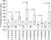
There was a trend toward decreased anteroposterior translation with the intramedullary reconstruction and increased anteroposterior translation with the extramedullary reconstruction compared to intact; however, this was not statistically significant (Table 1). There were also no statistically significant differences for the superoinferior translation comparing the intact specimens to either the intramedullary or extramedullary reconstructions (Table 2).
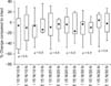
To compare the two reconstructions to each other, the percent change in translation compared to intact was calculated. The intramedullary technique provided significantly greater anteroposterior translational stability for four of the six loading conditions compared to the extramedullary reconstruction (Fig. 4). There was no significant difference in superoinferior translational stability between the two reconstructed specimens (Fig. 5).
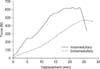
Representative load-displacement curves for both reconstruction techniques are shown in Fig. 6. The intramedullary technique also demonstrated improved load to failure characteristics including stiffness, yield and ultimate load to failure, deformation, and energy absorbed (Table 3). However, only two parameters were statistically different between the two reconstructions. The intramedullary reconstruction had a lower deformation at yield and a higher ultimate load than the extramedullary reconstruction.
The mode of failure for the intramedullary specimens was four Fibertape suture pullouts through the acromion and two clavicle fractures. For the extramedullary specimens, there were three coracoid fractures, one graft rupture, and two clavicle fractures.
DISCUSSION
The intramedullary free-tissue graft reconstruction of the AC joint was biomechanically stronger and provided increased anterior to posterior stability than the extramedullary reconstruction. There are several potential reasons to explain this finding. The intramedullary technique utilizes a more robust graft, generally 6-7 mm thick, versus 4.5 mm thick for the extramedullary technique, which is limited by the drill hole size. In addition, the intramedullary reconstruction is performed with Fibertape, allowing a thicker, stronger suture enhancement tied in a cruciate fashion superior to the AC joint, providing additional stability. Furthermore, the extramedullary technique only recreates the superior AC capsule, whereas the intramedullary technique essentially reconstructs the entire ligament. As previous studies have shown, the inferior capsule provides anterior stability, while the superior and posterior capsule serves as the major posterior restraint.17,18)
Similar to previous studies AC joint translation decreased as AC joint compression increased, indicating the importance of preserving the distal clavicle for improved stability.24,27) There were no significant differences in the superoinferior translational values between the two reconstructed specimens. Whereas, both techniques recreate a CC complex, the extramedullary technique utilizes drill holes in the clavicle and coracoid. As a result, several specimens failed via coracoid or clavicle fracture. Clavicle fracture following CC reconstruction using clavicular bone tunnels is a known complication.28) Contrary to our findings the primary mode of failure described by Grutter and Petersen20) was graft rupture in the CC loop portion with only one specimen failing by coracoid fracture. Our technique avoids drill holes in the clavicle and coracoid, therefore preventing the formation of stress risers and potential fracture.
The load to failure characteristics demonstrated the intramedullary technique had a statistically significantly higher ultimate load than the extramedullary technique; however, we were unable to reproduce the values reported by Grutter and Petersen20) for the extrmedullary reconstruction. The ultimate load to failure, elongation at failure, and stiffness for the specimens reconstructed with the extramedullary technique was 355 ± 123 N, 21.8 ± 7.4 mm, and 29.2 ± 12.5 N/mm in our study versus 774 ± 69 N, 14.45 ± 4.66 mm, and 59 ± 21 N/mm in the original study. This may be explained by differences in load to failure technique. We distracted the clavicle superiorly at a constant rate of 50 mm/min until failure, while they loaded at a rate of 10 N/sec until failure. Furthermore, Grutter and Petersen20) loaded the same specimen to failure four times, including the native AC joint complex first and then after three different reconstruction techniques which may have affected their load to failure testing values.
There were some slight differences in technique between our extramedullary reconstruction and the one originally described. In preparing the graft, we used a #2 Fiberwire suture as opposed to a #5 Ethibond used by Grutter and Petersen.20) They had one specimen fail due to suture rupture, thus we attempted to enhance the construct by using a biomechanically stronger suture. The second difference was that we used semitendinosis grafts as opposed to flexor carpi radialis (FCR) grafts. The reason they chose FCR was that it was readily available, however, they conceded semitendinosis or gracilis grafts would be a better choice for several reasons. First, the hamstrings are more likely to be used in clinical practice. Second, they have been shown to have increased strength as opposed to FCR, with the semitendinosis failing at 1,216 N.26) Finally, the authors had issues with inadequate length of the FCR tendon, thus supporting semitendinosis grafts as having abundant length for their anatomic AC complex reconstruction. Thus, we chose to use semitendinosis over FCR for our extramedullary reconstruction technique.
We have previously shown that this intramedullary technique of reconstructing the AC ligament restores native AC translational stability and that it is biomechanically superior to the popularly performed modified Weaver-Dunn procedure.23,25) This current study has demonstrated this technique to be biomechanically stronger and translationally more stable in the anteroposterior direction than the only other biomechanically tested technique described in the literature which anatomically reconstructs both the AC and CC ligaments. These authors, however, did not test translational stability, but rather focused on load to failure characteristics. While some studies reconstructed or repaired both the AC and CC ligaments, they did not biomechanically test them.21,29) Finally, in the biomechanical studies to date, testing has been done on reconstructed AC ligaments, ignoring the CC ligaments and vice versa.3,4,6,7,9,30)
Several limitations exist for the current study. The first limitation is that this is a cadaveric, time-zero study that only evaluates the repair immediately after the time of reconstruction. With the model, the effect of healing cannot be evaluated; clinical studies are needed to evaluate the clinical outcome of this technique. This model is a simplified cadaveric model incorporating an externally applied load to simulate AC joint compression and muscle loading was not evaluated in this study. However, since the amount of AC joint compression is unknown and can vary based on activity different AC joint compression loads were simulated.
In summary, the intramedullary free-tissue graft technique of reconstructing the AC ligaments, in combination with a CC sling graft reconstruction, provided greater anteroposterior stability and improved biomechanical strength than a previously described extramedullary technique. This technique has biomechanically proven itself in several studies, however no clinical studies have been undertaken to date. Future directions would include demonstrating clinical outcomes of this technique and comparing clinical results to other techniques in prospective, randomized studies.


 XML Download