

 PDF
PDF ePub
ePub Citation
 Print
Print


The periprosthetic fractures around the knee are becoming more common with the ever-increasing incidence of the total knee replacements.1) Most of the fractures are the low-velocity fractures, which occur as a result of a combination of the torsional and axial loads.2) Osteoporosis, local osteolysis, inflammatory arthritis and the revision knee arthroplasty may serve as the additional risk factors.3,4) Distal femur fractures are the most common compared to the fractures of the proximal tibia and patella.5,6) The surgical management of these fractures can be broadly classified into the attempts at osteosynthesis and the revision arthroplasty with or without a use of the allografts. Nonoperative treatment is indicated only in the undisplaced fractures in the low-demand individuals and the patients who are unfit for surgery.7) Osteosynthesis of the periprosthetic distal femur fractures pose several challenges because of the compromised bone quality and the non-feasibility of using the standard methods of fracture stabilization. Surgical management of these fractures can be associated with the high nonunion and complication rates.8,9)
The treatment options may have to be individualized based on the fracture site, displacement and the stability of the prosthesis used, with the classification systems to guide the surgical management. This retrospective study analyzes the outcomes of the distal femur periprosthetic fractures following TKA treated by the locked plating.
METHODS
From 2005 to 2009, 31 patients were admitted with the periprosthetic fractures following a total knee arthroplasty (TKA) at our institution. All records were retrieved from the hospital's prospectively maintained arthroplasty database. The Institutional Review Board approved the study. Periprosthetic distal femur fractures following a primary TKA treated by the locked plate osteosynthesis were included. Of the 21 patients satisfying the inclusion criteria, 20 patients who consented for the treatment were included in the study. There were 2 type I fractures and 18 type II fractures of the distal femur according to the classification of Rorabeck and Taylor.10) The mode of injury was a low-velocity fall in all patients.
The mean age was 73 ± 5 years. Fourteen were female and 6 were male patients. The mean duration between the arthroplasty procedure and the fracture occurrence was 20.8 ± 10.4 months. The mean follow-up was 39 ± 10 months.
Treatment Methods
The lateral vastus splitting approach was used, and the fractures were stabilized with a 4.5 mm precontoured distal femur locked plate (Synthes, Gurgaon, India). If the satisfactory fracture reduction and the limb alignment in the sagital and the coronal planes could be achieved closed, the minimal access plating technique was used. Failure to achieve a satisfactory reduction necessitated an open approach. The patients were kept non-weight-bearing after the surgery and progressed to full weight-bearing with the fracture consolidation. The range of motion exercises were encouraged immediately post-surgery to minimize motion loss.
Risk Factors
Osteoporosis (T score > -2.5) was evident in 13 patients. The bone quality was graded as osteopenia (T score, between -1 and -2.5) in the remaining 7 patients. Other risk factors included a grade II notching of the anterior femoral cortex in 1 patient.
Statistics
Continuous variables were expressed as mean and standard deviation. Categorical variables were presented as absolute and relative frequencies. The prefracture and follow-up functional outcome measures (Western Ontario and McMaster Universities Arthritis Index [WOMAC] and range of motion [ROM]) were compared using Wilcoxon analysis, and the level of significance was set at 2-sided p-value < 0.05.
RESULTS
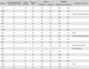
Refer to Table 1 for the patient and fracture demographics and the study outcomes.
Radiological Outcome
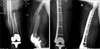
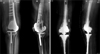
One patient died due to an unrelated illness and was lost to follow-up. Nineteen patients were available for the final analysis. Successful osseous union was achieved in 16 patients (84%) at the mean time of 22 ± 5 weeks (Fig. 1). The delay in union (lack of satisfactory progression at 4 months) was seen in 3 patients and was expedited with the iliac crest grafting. Union was ultimately achieved in 2 of the 3 patients by 8 months. Nonunion was diagnosed after a failed grafting in 1 patient (5%) with a deficient bone stock after 8 months (Fig. 2). She ultimately underwent a revision arthroplasty 11 months after the fracture. No patient had a significant deformity in the coronal or sagital plane (> 10°) postsurgery.
Functional Outcome
The mean ROM significantly decreased to 106° ± 12.8° at the last follow-up compared to 125° ± 9.1° before the fracture occurrence (p = 0.00). A similar trend was seen in the WOMAC scores, which decreased to 75.8 ± 4.2 compared to the prefracture score of 84 ± 3 (p = 0.00). At the last follow-up, 8 patients (42%) were community ambulant without support; 8 (42%) were community ambulant with the assistive aids; and 3 (16%) were restricted to home activities. At the last follow-up, 7 patients were fully satisfied with the outcome; 9 patients were somewhat satisfied; and 3 patients were not satisfied.
Complications
For the complications, six secondary procedures were carried out in 5 (26%) patients; the autologous iliac crest grafting in 3 patients; revision arthroplasty in 1 patient; and the knee manipulation under anesthesia in 2 patients after the fracture union to improve ROM. No deep infections were seen. None of the patients died in the early postoperative period (< 1 month). One patient developed basal atelectasis and a collapsed lung, and 1 patient developed acute renal failure in the perioperative period. Both patients recovered uneventfully.
DISCUSSION
Distal femur is the most common site for the periprosthetic fracture following TKA. The reported fracture risks following a primary TKA is 0.6% compared to 1.7% for a revision TKA.11) The goal of the management is to return the patient to the preinjury activity levels as early as possible. The surgical treatment is currently favored with the improvements in the implant design and surgical techniques. Conservative treatments may be associated with high rates of malunion, pain and stiffness. As such, such treatments should be reserved for the stable fractures in the low-demand and co-morbid individuals.12)
The locked plating and retrograde nailing are the currently favored techniques in treating the periprosthetic distal femur fractures. Several authors have reported successful results with the retrograde nailing.13,14) The technique is minimally invasive and biological but with a high incidence of malalignment, and the lesser union rates have been reported by some authors. Chettiar et al.15) reported the successful union in all 13 patients treated by the retrograde nailing in their study, though the axial malalignments in either the coronal or the sagital plane (> 5°) were seen in 30% of the patients. Similarly, Large et al.16) reported either a nonunion or malunion in all 7 patients treated with the retrograde nailing in their series.
Significant coronal malalignments were not seen in our study. Rotational alignment is difficult to appreciate intraoperatively. We compared the position of the lesser trochanter with the opposite limb to roughly establish the right rotation.17) The rotation of the hip was also compared with the opposite hip after fixation. While several methods have been described to assess the rotational alignment during the surgery, none of the methods are reliable. The rotational malalignments, though less common, have been described with the plating, especially with the minimal access techniques.18)
The locked plating can be minimally invasive, and the successful healing rates have been reported even in the osteoporotic bones with the lesser incidence of malalignment.19,20) Most of the fractures in our study were treated with the minimal access locked plating, which preserved the fracture biology and minimized the muscle damage. Despite the limitations in the bone quality, we were able to achieve the satisfactory union rates. The study outcomes in terms of the union rate, function and incidence of complications were comparable to the previously published studies.21,22) Osteoporosis is a major determinant in the fracture occurrence following arthroplasty.23) It can also complicate the fixation strategies and delay rehabilitation. Sixty-five percent of the patients in the current study suffered the osteoporotic insufficiency fracture. Long-term medical management of osteoporosis may be justified after the arthroplasty procedure, especially in females.
Despite the satisfactory rate of union with an acceptable complication rate, only around 45% of the patients were fully satisfied with the treatment and were able to return to the pre-fracture activity levels. There was a significant drop in the functional level as evident from the WOMAC scores and the range of motion analysis. Additional risk factors including osteoporosis, increasing age and medical comorbidities can prolong rehabilitation and may contribute to the deterioration in function. The retrospective analysis, a relatively small sample size and the short follow-up are the limitations of this study. All data used in the study were collected from a prospectively maintained arthroplasty database. In conclusion, an acceptable to satisfactory clinical results can be achieved with the locked plate osteosynthesis for the periprosthetic distal femur fractures following TKA.
 XML Download