

 PDF
PDF ePub
ePub Citation
 Print
Print


Locking plate constructs have shown promising results for treating displaced and unstable proximal humerus fractures,1-3) but the use of the this technique has not been without complications.3-5) Studies that have evaluated the outcomes of patients treated with locking plates for proximal humerus fractures have shown that one of the most frequent complications of this technique is intra-articular penetration of the locking screw.3-5) No study has described the superiority of longer versus shorter locking screws placed into the humeral head for fixation of these fractures. This article describes a variation of the locking plate application that utilizes short locking screws in the humeral head in conjunction with suture fixation to the rotator cuff as a means of minimizing the potential for intra-articular screw penetration and increasing stability.
TECHNIQUE
The patient can be positioned supine on a radiolucent operating room table or placed in the beach chair position. An image intensifier is positioned at the head of the bed. Fluoroscopic images are obtained to confirm visualization with the image intensifier prior to prepping and draping the patient's shoulder, neck, and arm. Full anesthetic relaxation allows for less traumatic retraction of the deltoid and minimizes dynamic forces on the fracture fragments during reduction.
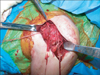
A deltopectoral exposure is used to expose the proximal humerus, as described previously by Badman and Mighell.6) The coracoacromial ligament may be partially or completely released. Similarly, the coracohumeral ligament is released. The long head of the biceps brachii tendon is identified at its position medial to insertion of the pectoralis major on the humerus. The pectoralis does not typically need to be released. However, if left in situ, the long head of the biceps brachii can be a source of pain and we often tenodese it at the time of plate fixation in older patients or those with grossly poor tendon quality. The transverse humeral ligament is released with a knife or electrocautery as the biceps is traced superiorly, and as the tendon courses superiorly, it is used to define the rotator cuff interval. After the rotator interval is released to the base of the coracoid process, the long head of the biceps can be released from the supraglenoid tubercle and superior glenoid labrum if there is a plan for tenodesis. Heavy nonabsorbable sutures are placed in the subscapularis, supraspinatus, and infraspinatus tendons at the myotendinous junction. Temporary traction sutures are often necessary to help mobilize the tendons to obtain better suture purchase more medially. The bone quality in these patients is typically poor, and sutures should be placed in the stronger rotator cuff tendons rather than through the soft, metaphyseal bone of the tuberosities. We have found that unlocked horizontal mattress sutures are adequate. Traction sutures should be placed in the tendinous insertions to hold and reduce the fragments securely to the plate (Fig. 1).
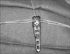
A low-profile, precontoured, peri-articular locking plate with angular stable screws and suture eyelets is selected to provide fracture fixation. Separate sutures are passed through the plate eyelets prior to applying the plate (Fig. 2). Ideally, a superior suture is placed for the supraspinatus tendon, an anterior suture for the subscapularis, and a posterior suture for the infraspinatus tendon. The plate is applied to the proximal humerus lateral to the biceps tendon. Care should be taken to avoid medial dissection, which entails detaching the pectoralis major tendon, and risks injury to the posterior humeral circumflex artery.
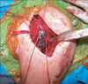
A provisional reduction of the surgical neck can be held with Kirschner wires and confirmed with an image intensifier and by direct inspection. The tuberosities are reduced via the traction sutures with minimal manipulation of the metaphysis to prevent further fracture comminution. The plate is secured to the humeral head and/or the shaft using Kirschner wires. The initial screw should be diaphyseal, bicortical, and non-locking. This allows compression of the plate against the humeral shaft and allows subsequent reduction of the tuberosities to the shaft via the plate (Fig. 3). The plate can be moved caudal or cephalad as needed using the oblong hole in the plate. It is critical not to reduce a fracture in internal rotation, as this will limit the patient's ability to regain functional external rotation postoperatively. To ensure that this does not occur, reduction and plating are performed with the arm in 30° of external rotation. The position of the plate is then evaluated using fluoroscopy to ensure appropriate placement such that the plate is not too proximal to impinge on the coracoacromial arch with shoulder abduction. Similarly, the plate should not be positioned too distal such that fixation into the head is limited.
Reduction techniques are employed to reduce the head to the shaft by using the sutures in the rotator cuff to gain control of the head. Alternatively, the humeral head can be reduced or stabilized by manipulation with blunt elevators or joysticks such as Kirschner wires or Schanz pins. The use of elevators or joysticks can be problematic when working with osteoporotic bone, due to poor bone quality and further fragmentation at the site of application of these reduction tools.
Once reduced, fixation into the head is limited to five or more short (32-38 mm), fixed angle screws augmented with suture fixation of the rotator cuff (which has control of the head) directly to the plate. Screw length is based on the overall size of the proximal humerus. Once a single screw is placed and its length is confirmed by an image intensifier to be > 1-2 cm from the articular surface, all other holes are drilled and screws of the same length are placed. This is done to prevent cut-out and intra-articular penetration of the screws. In addition, this can lead to decreased operative time. If the long head of the biceps tendon was released, it can be tenodesed to the pectoralis major and the rotator cuff interval prior to a layered closure.
Postoperative Rehabilitation
Patients are placed in a sling for comfort only and are encouraged to perform non-load bearing activities and pendulum exercises immediately after surgery. If there are no signs of reduction loss, patients are increased to full weight-bearing with the arm over the course of 4-6 weeks. All restrictions are lifted at 12 weeks if radiographs reveal healing.
DISCUSSION
Proximal humerus fractures account for 4-5% of all fractures.7) The majority of these fractures occur in the elderly population, particularly in those with osteoporosis. Approximately 85% of proximal humerus fractures are minimally displaced or stable and can be successfully treated with conservative management and early motion.8-10) The remaining 15% of fractures are displaced or unstable and require surgical intervention because of poor results with non-operative treatment.9) Plate and screw fixation offer the best chance for stable fixation of multi-fragmented fractures. Importantly, no study has described the biomechanical superiority of long versus short screws placed in the humeral head for fixation of these fractures.
Locking plate constructs have shown promising results for treating displaced and unstable proximal humerus fractures,1-3) but the use of this technique has not been without complications.3-5) Studies evaluating the outcomes of patients treated with locking plates for proximal humerus fractures have shown that one of the most frequent complications (16-23%) of this technique is intra-articular penetration of the locking screw.3-5) It was noted that this complication is more common in patients > 60 years in whom osteoporotic bone is more likely to be found. We believe that the concept of subchondral screw fixation (as in load bearing joint periarticular fractures such as femoral neck fractures) is a misuse of the locking design for proximal humerus fractures in which rotator cuff tissue integrity often exceeds that of the metaphyseal bone of the humeral tuberosities. For this reason, we use short, divergent locking screws and suture fixation to minimize the risk of varus malunion, plate failure, and intra-articular screw penetration. We have treated 53 proximal humerus fractures at our institution with this fixation technique. None have had intra-articular screw penetration or cut-out and only two patients had an asymptomatic varus malunion at an average follow-up of 16 months. It is our belief that such a technique reduces the incidence of screw penetration into the glenohumeral joint and provides stable fixation for healing. Further biomechanical and long-term clinical data are necessary to substantiate these hypotheses.
 XML Download