

 PDF
PDF ePub
ePub Citation
 Print
Print


Many options are available to manage patellae during revision total knee arthroplasty (TKA). After the decision to revise has been made, a treatment option is chosen based on the condition of the patella before and after implant removal. If a conventional onlay-type prosthesis is used, a minimum of 8-12 mm of residual patella is required to obtain adequate support for fixation.1) However, decision-making regarding resurfacing options is not straightforward if less than 8 mm of bone stock remains.
Recently, new implants have been introduced for patellar resurfacing, such as, the biconvex prosthesis or the trabecular metal patella.2-4) But, the use of these implants is arguable because definite indications have not been established and information on long-term follow-up results is sparse. Therefore, we studied a resurfacing technique conducted with widely used instruments, and here, we introduce this technique which is based on the use of a conventional onlay-type prosthesis with cementing and transcortical wiring, and which although straightforward, enables firm fixation.
TECHNIQUE
Between May 2009 and August 2010, 30 consecutive revision knee arthroplasties were performed. The procedure included augmentation with transcortical wiring of an onlay-type prosthesis in twelve knees (nine patients) after it had been determined that conventional resurfacing was inadequate. This technique was indicated when the thickness of the remnant patella was less than 8 mm and the cortical rim was not intact. There were 9 patients (12 knees) with a mean age of 68 years (range, 53 to 80 years) at the time of surgery. Thickness of the remnant patella ranged from 3.2 to 7 mm. The patellae were revised because of infection (five knees), loosening of the patellar component (four knees) and polyethylene wear in the presence of a metal-backed patella (three knees). An onlay-type, all polyethylene prostheses (Universal Dome Patella, Scorpio, Stryker, NJ, USA) which are compatible with the femoral component were used. All patellar prostheses were fixed with acrylic bone cement and 24 gauge steel wires.
Surgical Technique
After arthrotomy using a medial parapatellar approach, the patella is everted laterally and fixed in this posture by grasping the quadriceps tendon or patella tendon with towel clips. Cement stuck to the undersurface of the prosthesis is meticulously removed with an oscillating saw and a high-speed burr after implant removal. The new, properly sized patellar prosthesis is then placed on the undersurface, implant location is decided and the margin of the prosthesis and the contact points for each peg are marked. Bone bed preparation is performed with a burr according to marking lines and contact points. On fitting the patellar prosthesis, a check is made to determine whether it matches the concave portion of the undersurface and confirm the peg sites. Three points are then drilled for the pegs and bone holes are placed for wire passage.
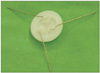
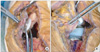
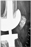
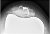
The next steps involve connecting the wires to the pegs and passing through drill holes. A wire is connected to one peg by winding it around the narrow part of the neck under the peg head and fixing it to the peg. The wire is then twisted like a two-stranded rope (Fig. 1). The same procedure is performed at the other two pegs. The three wires on the pegs are passed from the undersurface of the patella to the outer surface through the bone holes. Before the prosthesis is brought into contact with the patella, the space between the bone bed and the polyethylene is filled with cement (Fig. 2). After approximating the patellar prosthesis to remaining bone as closely as possible, the prosthesis is compressed with a compressor and the cement is given time to harden. Subsequently, sufficient tension is placed on the three wires on the outer surface with wire holders and the wires are twisted together immediately above the cortex. After checking that the wires are under sufficient tension, they are cut and their ends are pressed with an impactor to prevent irritation (Fig. 3). Radiographs are checked immediately after surgery (Figs. 4 and 5). The average thickness of the patella after surgery was 14.6 mm.
DISCUSSION
If the patellar component can be retained during revision TKA, this may offer the best option in terms of morbidity. However, if the prosthesis is malpositioned, damaged or mechanically loose, removal is probably the advisable option.
The onlay-type all polyethylene patellar prosthesis is an efficient tool when patellar bone stock is sufficient, being over 10 to 12 mm.5) But it is not easy to determine an optimal procedure if bone stock is poor. According to Maheshwer et al.,2) patellar resurfacing using an all polyethylene biconvex patellar prosthesis is successful when the remnant patellar thickness is less than 10 mm and the peripheral rim is intact.
When the bone stock is too thin or the peripheral rim has been destroyed, it is highly likely to fail when a prosthesis is only cemented to bone. A biconvex patellar prosthesis may also not be likely to be effective. In our series, the remnant patellar thickness averaged 5.6 mm (range, 3.2 to 7 mm) and variable amounts of the peripheral patellar rim were damaged. With regard to these cases, some authors advocated alternative options including a porous tantalum component, a bone grafting and soft tissue flap procedure or a gull-wing osteotomy. Nelson et al.4) evaluated short-term results following patellar resurfacing with a trabecular metal patella in the setting of marked patellar bone loss. The results were good or excellent in 17 of 20 patients. Hanssen6) suggested a bone grafting and soft tissue flap procedure. At final follow-up of nine patients, it was found that the mean patellar thickness increased from 8 to19.7 mm and their functional outcomes were improved. Klein et al.7) performed a gull-wing osteotomy in a nonresurfacable patella and reported that there was a significant improvement in the range of motion and Knee Society scores. Though several shortcomings were encountered during follow-up period, the above techniques showed reasonable outcomes.3,4,6,7) However, further follow-up is required to assess the longer term benefits of these methods.
Our technique involves cementing and cortical wiring of an onlay-type prosthesis. The deficient patella was revised using this technique when the thickness of the remnant patella was less than 8 mm and the cortical rim was not intact. This procedure has an advantage of using proven and unsophisticated instruments. Moreover, it is easily reproducible and does not need a long learning curve, in our experience. Because it protects the extensor mechanism and provides the same contact during patellofemoral articulation as primary TKA, painless articulation is possible and hereby, the range of motion is improved. However, there are a few concerns about the use of this procedure in thin patellae. It may not be indicated if the osseous cortical shell is too thin for wiring, so the use of transcortical wires place the patella at risk to fracturing. Also, the problems related with prominent hardware also may occur as is oft en seen with internal fixation of patellar fractures. At a mean 10.3 months follow-up of our series, there was one patellar fracture arising during flexion exercise one week after surgery that was treated with partial patellectomy. There were no complications associated with hardware. Further follow-up is being continued to investigate the surgical outcomes.
Our described technique includes connecting wires to the three pegs of the patellar component, passing the wires from the undersurface of the patella to the outer surface through the bone holes and, after compression of a cemented prosthesis, twisting the wires under sufficient tension. We believe that this technique provides a good alternative option in managing the deficient patella in revision total knee arthroplasty, although any prolonged benefits will require long-term follow-up.

 XML Download