

 PDF
PDF ePub
ePub Citation
 Print
Print


Proper ligament balancing, restoration of the mechanical axis and component alignment are essential for the success and longevity of a prosthesis.1-3) Physiological recovery of the leg alignment is the most closely related factor. Acceptable tibiofemoral alignment is difficult to obtain by the eye because of a tourniquet, drapes and subcutaneous fat. External jigs or intramedullary rods are needed. Computer-assisted navigation offers considerable improvement for more reliability, achieving lower extremity alignment than a conventional instrumented total knee arthroplasty (TKA) technique. Moreover, computer-aided knee arthroplasty is beneficial for accurate alignment in several complex situations, such as posttraumatic femoral deformity and retained femoral hardware, which cannot be applied by traditional instrumentation.
This paper reports 2 successful cases of navigation-assisted TKA for severe osteoarthritis of the knee with retained hardware of the distal femur.
CASE REPORTS
Case 1
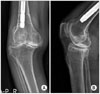
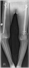
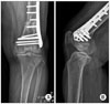
A 71-year-old woman presented with pain of the right knee with 2 year duration. She had been involved in a slip down and sustained right femur shaft fracture 3 years earlier, as well as a left femur shaft fracture 5 years ago. Preoperative radiographs of the right knee in the anteroposterior and lateral views showed tricompartment osteoarthritis and a retained intramedullary nail. The femoro-tibial angle that formed between the mechanical axis of the femur and tibia axis was 25° varus (Figs. 1 and 2).
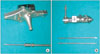
The patient underwent a right TKA with computer navigation using the Stryker 4.0 system (Stryker Co., Allendale, NJ, USA), cemented, and posterior cruciate ligament sacrificing type (Scorpio, Stryker Co.,). A midline skin incision was made. A 6.5 mm bicortical self-tapping anchoring screw was inserted 10 cm below the tibiofemoral joint line. Another type anchoring pin for the femur was used to avoid a collision with the intramedullary nail. This type of anchoring pin is smaller (3.0 mm) than a anchoring screw, and unicortical anchoring is available (Fig. 3). Two 3.0 mm self-tapping, anchoring pins were inserted into the femur. A femur and tibia resection were performed according to the light-emitting diode tracker of a navigation system and cutting jig. There was a severe bone defect on the medial condyle of tibia. Therefore, tibial side arthroplasty was performed with a stem and medial metal augmentation. The ligamentous release and balance were also performed based on a trial reduction of the prosthesis and guide in a navigation system. The femoral and tibial components were implanted with cement. The patella was not resurfaced. The knee was irrigated and the wound was closed over the suction drain.
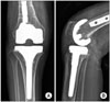
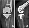
Postoperative radiographs revealed good alignment of the prosthesis. Both components were centered to the midline of the joint, and the overall knee alignment was 5° valgus (Fig. 4). After TKA, the patient could walk with partial weight bearing and full weight bearing 1 day and 7 days after surgery respectively.
Case 2
A 79-year-old woman presented with pain of the left knee with a 3 year duration. The patient was involved in a traffic accident 15 years earlier and sustained a left distal femur fracture. The fracture was treated with a plate and screw fixation.
Preoperative radiographs of the left knee in the anteroposterior
and lateral views revealed tricompartment osteoarthritis and a containing plate and screws on lateral side of the femur (Fig. 5). The femoro-tibial angle that formed between the mechanical axis of the femur and tibia axis was 15° varus. A left knee CT scan was performed to determine the position of the screws.
The patient underwent right TKA performed with computer navigation using a Stryker 4.0 system, cemented, posterior cruciate ligament sacrificing type (Scorpio). A midline skin incision was performed. A 6.5 mm bicortical self-tapping anchoring screw was inserted 10 cm below the tibiofemoral joint line. Another type of anchoring pin was used on the femur to avoid a collision with the screws. The pins were smaller (3 mm) and unicortical anchoring was available. Two 3.0 mm self-tapping, anchoring pins were inserted into the femur. The lateral epicondyle, which was covered with a plate, could not be checked. A multiple checking system, Whiteside's line, posterior condylar line and tibial cutting plane, were adapted. A femur and tibia resection were performed depending on the light-emitting diode tracker of the navigation system and cutting jig. The most distal screw impinged on the box cutting for the posterior cruciate ligament sacrificing type and one screw was removed. Ligamentous release and balance were also performed based on a trial reduction of the prosthesis and guide in the navigation system. The femoral and tibial components were implanted with cement. The patella was not resurfaced. The knee was irrigated, and the wound was closed over the suction drain.
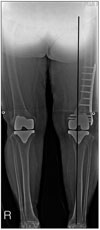
Postoperative X-rays revealed good alignment of the prosthesis. Both components were centered to the midline of the joint, and overall knee alignment was 6° valgus (Figs. 6 and 7). After TKA, the patient could walk with partial weight bearing and full weight bearing 1 day and 7 days after surgery respectively.
DISCUSSION
A navigation system has been reported to improve the accuracy of bony cuts and restore the mechanical axis in TKA.4-6) Computer-assisted TKA using a surgical navigation system provides accurate cuts of the distal femur and proximal tibia without the need for intramedullary instrumentation. Intramedullary instruments cannot be used in patients with previous trauma and substantial residual bony deformities, or retained hardware that cannot be removed.
In old age, bone is usually osteoporotic and the fractured bone fixed with an implant may be more osteoporotic. In conventional TKA, when removing the implant, the space where the implant had been removed can be stress riser for a given load after TKA.7,8) Early ambulation and early continuous passive motion should be performed more carefully. In these 2 cases, partial weight bearing walking and full weight bearing walking was possible 1 day and 7 days after surgery, respectively. The patients received continuous passive motion 1 day postoperatively.
A different type pin was used at the femur. The pins were smaller (3.0 mm) than used at the tibia and could be anchored unicortically. The reasons for changing the anchoring screw to pins were to reduce the anecdotal fracture and collision with retained hardware. However, a unicortical pin at the metaphysis for the tracker often has insecure fixation that can cause poor registration during navigation-assisted TKA. Two anchoring pins were inserted for the femur but three pins can be inserted for more secure fixation. Some cases with an anecdotal fractures have been reported after the placement of navigation tracker pins.9,10) There are a few reports that a smaller size and unicortical fixation pin is safer. In one anatomic study, the findings indicated the close proximity of the popliteal vessel to potential injury with the bicortical placement of tracker pins in the anteroposterior direction in a typical total knee approach. In particular, the neurovascular route may be distorted in a healed femoral fracture. Therefore, an attempt was made to anchor it unicortically. Two anchoring pins were inserted for secure fixation of the femur.
In conclusion, navigation-assisted TKA is feasible and less invasive for TKA of a retained implant of the distal femur after a fracture.
 XML Download