

 PDF
PDF ePub
ePub Citation
 Print
Print


Infective spondylitis, tuberculous infection, Crohn's disease, and diverticulitis are known causes of psoas abscess; however, a perforating carcinoma of the colon causing this condition, is rare.1,2) We report a case of psoas abscess in a middle aged male that is the result of a spontaneous rupture due to colon cancer.
CASE REPORT
A 44-year-old male was visited for back pain and mass around left buttock. He had a medical history that included a diagnosis of pulmonary tuberculosis, from which he had recovered completely 20 years previous. He was under medication from a local clinic and pharmacy for 1 month prior to visiting the hospital due to cold symptoms and lower back pain.
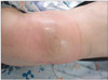
Vital signs on visit were blood pressure at 130/80 mmHg, heart rate at 72 beats/minute, respiratory rate at 18/minute and body temperature was 38.3℃. The mass was painful and palpated on the left buttock area, and is seen as a reddish skin color around the anterior superior iliac spine (Fig. 1).
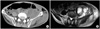
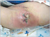
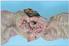
Laboratory investigations on admission were the following: white blood cell count was 16,660/mm3, hemoglobin was 10.5 g/dL, platelet count was 631,000/mm3, C-reactive protein was 21.9 mg/dL, and erythrocyte sedimentation rate was 120 mm/hour. Other biochemical tests and coagulation tests were normal. Abdominal computed tomography (CT) and magnetic resonance imaging (MRI) showed a large psoas abscess in retroperitoneal space and abscess on left lower quadrant anterior abdominal wall (Fig. 2). Based on a diagnosis of psoas abscess, the patient was given ciprofloxacin and metronidazole as an empirical injection, then an incision and drainage was performed. Ciprofloxacin and metronidazole were injected for an additional 7 days after this procedure, however Streptococcus agalactiae and Streptococcus aginosus were cultured. This finding prompted a change in treatment to 3rd generation cephalosporin and metronidazole. A skin defect, 4 × 3 cm in size, developed around left anterior superior iliac spine. Debridement, followed by a local flap, with superficial skin graft was performed. Patient was discharged after relief of symptoms. Ten days post discharge, the patient experienced fecal discharge from the local flap site (Fig. 3). The patient underwent an operation for enterocutaneous fistula, which was performed by the Department of General Surgery. During the surgical procedure, a tumor mass was found in the descending colon and was adherent to the retroperitoneum. The tumor had ruptured spontaneously and formed enterocutaneous fistula track, but there were no peritoneal seeding. A left hemicolectomy was performed (Fig. 4).
Histopathologic results of the resected colon mass confirmed a diagnosis of adenocarcinoma. The histopathologic results of the fistula track and capsule of abscess were granulation tissue with acute suppurative inflammation without tumor cell. There was no lymph node metastasis. The patient is currently undergoing postoperative chemotherapy and remains symptom-free.
DISCUSSION
Reported cases of psoas abscesses have been attributed to tuberculous diseases of the spine, infective spondylitis, Crohn's disease, and diverticulitis.3)
The incidence of perforated colon cancer ranges from 3% to 10%.4) Development of intra-abdominal abscess from spontaneous rupture of colon cancer is very rare, having an incidence of 0.3-0.4%.1,2) The psoas muscle is a retroperitoneal structure lying outside the endoabdominal fascia, such that it is protected from intra-abdominal muscle.5) Similar to this case, a psoas abscess from colon cancer usually has been preceded by perforation of cancer and commonly with fistula formation. Abdominal CT is useful in diagnosis and MRI helps in judgment of abscess location, determination of lesion boundaries, and the spread to surrounding soft tissue.
In this case, the patient was relieved of symptoms from the psoas abscess by routine incision, drainage and antibiotic therapy in this case. However, the wound culture from the operation was positive for S. agalactiae and S. aginosus which are unusual findings from this location. This led the authors to doubts about the original diagnosis of an uncomplicated psoas abscess and prompted the taking of abdominal CT and MRI.
We discovered that the patient's intra-abdomen was connected with retroperitoneum and fecal discharge, which were observed on the local flap. During the operation under enterocutaneous fistular impression, we found a ruptured tumor mass in the descending colon, which had adhered and connected to the retroperitoneum. The ruptured colonic mass had formed a fistula into the buttock area. The case report concerned a thigh abscess with perforated colon cancer; the route by which the abscess spread from abdominal sources were by 2 means in this case: one was the direct soft tissue extension of infection from the extraperitoneal portion of colorectum; the second was the extension of infection into the other site via naturally occurring defects in the abdominal wall.6) As previously described, perforated colon cancer is rare. Fistular formation is uncommon and occurs only in approximately 15% of all perforated colon cancers.7) In this case, abscess and fistula of perforated colon cancer were coexistent.
It must be recognized that ruptured colon cancer can be a rare cause of psoas abscess. If the result of wound culture is an uncommon strain or normal flora of bowel and the patients have continuous wound problem, surgeons should consider spontaneous rupture of colon cancer as part of the differential diagnosis in these cases.
 XML Download