

 PDF
PDF ePub
ePub Citation
 Print
Print


Primary spondylitis of a vertebral body rarely progresses to involve the posterior element.1) Less than 5% of the cases are known to involve only the posterior element. Most pyogenic vertebral infections involving the posterior element occur at the articular facets. There are only a few case reports of involvement of the pedicle, lamina and the spinous process.2-4)
To the best of our knowledge there has been no report of osteomyelitis involving the transverse process of the spine. Our report is the first report of osteomyelitis involving the transverse process of the spine.
CASE REPORT
A 45-year-old male, who was transferred from a local clinic, presented with a one month history of a swollen painful back. He had experienced intermittent back pain over the previous 2-year period. He denied a history of acupuncture or local injections. A mass-like swelling was palpated on the right side of his back. The range of motion of his back was slightly limited, but there was no radicular pain. The motor, sensory and deep tendon reflexes were normal in both lower extremities.
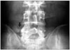
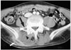
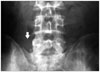
The white blood cell count and the erythrocyte sedimentation rate were 9,000/mm3 and 76 mm/hour, respectively. The plain radiographs indicated only subtle changes in the right transverse process of the 5th lumbar vertebra (Fig. 1). T2 weighted axial magnetic resonance imaging (MRI) revealed a space-occupying lesion, which was located behind the right paraspinal muscle, and it was connected to the paraspinal muscle (Fig. 2). Enhanced computed tomography (CT) revealed a 7 × 4 × 1 cm-sized space-occupying lesion with a rim enhancement pattern and swelling of the surrounding soft tissues. CT also revealed an osteolytic lesion (2 × 1 cm in size), which was located in the right transverse process of L5, and it was similar to Brodie's abscess in the long bones (Fig. 3).
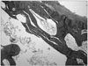
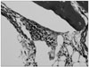
Open drainage of the abscess and a resection of the right transverse process of L5 were performed under general endotracheal anesthesia (Fig. 4). Grossly, the osteolytic lesion was surrounded by a sclerotic bony margin and there was pus in the abscess cavity, and the pus had gravitated anteriorly and posteriorly to the paraspinal muscle; this suggested chronic osteomyelitis. Th e pus culture demonstrated a mixed infection with Staphylococcus aureus and coagulase-negative staphylococcus. The histological examination revealed an infiltration of chronic inflammatory cells into the dense lamellar bone spaces, which was all consistent with chronic osteomyelitis (Figs. 5 and 6).
The patient received an intravenous injection of 1st generation cephalosporin (cefotiam hydrochloride 1.0 g bid) for 4 weeks, which resulted in the complete resolution of symptoms and a decrease in the erythrocyte sedimentation rate to normal and this lasted during follow-up. At the two year follow-up, the patient had normal spine functions.
DISCUSSION
Many spinal infections occur as a result of the hematogenous seeding of bacteria from various distant sites. Batson5) first proposed the valveless venous system as a route for the spread of a pelvic or genitourinary infection to the spine. Wiley and Trueta6) suggested the arterial network as the most likely source of bacterial spread to the spine. The transverse process contains end-arterioles and sinusoidal vessels that are similar to but less vascularized than the metaphyseal area of the vertebral body. An infecting organism enters the end-arterioles via the dorsal spinal plexus arising from a segmental artery. Once established, an infection within the transverse process can extend through the cortex into the paraspinal soft tissue.2,6)
Invasive diagnostic and treatment efforts have been associated with iatrogenic infections of the spine. In addition, direct bacterial inoculations of the spine via a penetrating trauma such as stab and gunshot wounds are other sources of infection. In our case, there was no evidence of any pelvic or genitourinary infections, nor was there any evidence of an iatrogenic infection and direct inoculation. Therefore, the arterial hematogenous route is the most likely mechanism of the osteomyelitis in this case.
Staphylococcus aureus is clearly the main organism in hematogenous vertebral osteomyelitis, and it accounts for more than 55% of all the reported cases. A polymicrobial cause is quite unusual and it accounts for less than 2.5% of the cases.7) In our case, 2 staphylococcus species, Staphylococcus aureus and coagulase-negative staphylococcus were identified. Our patient denied a history of acupuncture or local injections. But one may consider the possibility of iatrogenic infection.
Imaging studies are important for the diagnosis, the treatment and its planning, and for monitoring the treatment outcome of a spinal infection. The sensitivity of CT is high, but it lacks specificity. MRI is sensitive, specific and accurate and it is the method of choice for examining spinal infections.8) In our case the osteolytic lesion was not detected by MRI, while an accurate diagnosis was made by CT. Therefore, we believe that CT is more sensitive to the subtle changes in the bone than MRI, and MRI combined with CT is a more useful modality for evaluating transverse process osteomyelitis.
The treatment modality is determined by the extent of the infection and the degree of neurological compromise. In our case, the paraspinal abscess was surgically drained, and the primary osteolytic lesion was resected in order to eradicate the infection focus. Although 6 to 8 weeks of antimicrobial therapy is the generally accepted regimen, a recent report recommended 4 weeks of an appropriate high-dose, parenteral antimicrobial therapy,7) and this was adopted in our case.
King and Mayo9) reported that subacute osteomyelitis of the spine is a subtype of subacute hematogenous osteomyelitis. However, they did not introduce transverse process osteomyelitis.
Transverse process osteomyelitis is extremely rare, and this can result in a misdiagnosis or a delayed diagnosis. This report shows the transverse process is a possible location of vertebral osteomyelitis, and transverse process osteomyelitis with paraspinal abscess is an unusual cause of back pain. Therefore, transverse process osteomyelitis should be borne in mind as part of the differential diagnosis when conducting an examination of back pain.

 XML Download