

 PDF
PDF ePub
ePub Citation
 Print
Print


Although many radiologic parameters, such as the acetabular index (AI),1-4) the center-edge angle (CEA),5) the center-head distance discrepancy (CHDD),6) Smith's c/b and h/b ratios,7) and the amount of femoral head coverage (HC),8) have been proposed to indicate the degree of hip dysplasia, none of the radiologic parameters (either singly or in combination) accurately predicts acetabular development in all cases. For example, Chen et al.6) reported that 96% of hips with a CHDD ≤ 6% had satisfactory results, whereas hips with a CHDD > 6% had 78% unsatisfactory results. Similarly, Kim et al.9) divided patients into four groups based on the CHDD values and the orientation of the sourcil, and reported satisfactory results in 19 of 20 hips (95%) with a CHDD < 6% and horizontal sourcils, while 4 of 5 hips (80%) with a CHDD < 6% and upward sourcils had unsatisfactory results; 8 of 9 hips (88.9%) with upward-oriented sourcils had unsatisfactory results. These studies confirm that while CHDD and AI are important predictors of residual dysplasia, CHDD and AI fail to account for up to 20% of outcomes. Additionally, the often-stated goal of "concentric reduction" of the hip has never been precisely defined.
To overcome these shortcomings of radiograph-based management of hip dysplasia, it seems reasonable to look to soft tissue and/or cartilaginous structures for clues to other avenues of treatment. As the labrum is well-known to have an important role in the growth of the acetabulum,10-17) it is an obvious place to begin. Therefore, we initiated a study using MR imaging of dysplastic hips with emphasis on the labrum to answer or clarify the following questions: 1) What is the usual range of labral angles in normal and dysplastic hips?, 2) What is meant by concentric reduction of the hips?, and 3) How can radiographic and MR-based parameters be used together in managing developmental dysplasia of the hip (DDH)?
METHODS
Twenty-five range of motion (ROM)-magnetic resonance imagings (MRIs) (one each for 25 patients) were selected for this analysis. ROM refers to range of motion because the images were taken in the following positions: neutral; abduction; abduction-internal rotation; abduction-internal rotation-flexion; and adduction. The criteria for selection of patients and MRI were: 1) unilateral hip dysplasia after closed reduction or open reduction without previous bony surgery in the femur and pelvis; and 2) a clear ROM-MRI, which showed the labrum, articular cartilage, and surrounding soft tissues in all five positions of both hips. All of the dysplastic hips studied had one or more of the following on radiographs: AI ≥ 25°; CHDD ≥ 6%; subluxation; a widened medial or superior joint gap; or an upward-oriented lateral sourcil.9)
Before ROM-MRI, our 25 patients had 2 types of hip dysplasia (dislocation, 21 hips; subluxation, 4 hips). Fifteen hips received non-surgical treatment (Pavlik harness, 2 hips; abduction brace, 3; closed reduction, 10) and 10 hips were treated surgically (open reduction). All surgical procedures were performed at our institution by the senior author. Interposed ligamentum teres and soft tissues were excised and the labrum was preserved. Each patient also required two or three periods of hip spica cast immobilization after arthrographic evaluation. The mean patient age at the time of primary treatment was 10.0 months (range, 4 to 18 months) in the closed reduction group and 14.5 months in the open reduction group (range, 12 to 16 months), while the mean age at the time of MR imaging was 44.1 months (range, 22 to 88 months). There were 22 females and 3 males; involvement was on the left side in 18 and the right in 7.
Technique of ROM-MRI Scanning
All MRI scans were performed on a 1.5-T imaging unit (Magnetom Sonata; Siemens, Erlangen, Germany). Three consecutive T2-weighted coronal scans of the anterior, middle, and posterior parts of the hip joint were made in each of five positions: neutral; abduction; abduction-internal rotation; abduction-internal rotation-flexion; and adduction. Our reliance on T2-weighted scans reduced the conventional scanning time and minimized the time that young children had to remain in the MRI scanner. The scanning time for each position was approximately 4 minutes. In a T2-weighted image, the articular cartilage and the muscle have an intermediate signal intensity, while the labrum has low intensity and is triangular. Due to the abnormal accumulation of fluid in a subluxated hip, the widened joint space shown on the radiograph is an area of high intensity between the femoral head and the acetabulum.
Initially we used a specially designed foot holder which rested on the main operating MRI table and slid into the bore of the magnet. The foot holder consisted of a main frame with specially made shoes for the left and right feet. However, as the study progressed, it was found easier for the examiner to manually change the position of the patient's feet for each scan. Younger children were sedated, while explanation of the scanning procedure was helpful in obtaining the cooperation of older children without sedation.
In each case the abduction angle was made as large as possible, but it was limited by the size of the child and the 60-cm bore diameter of the machine; mean abduction angle was 22.7° (range, 9 to 44°). In adduction, the affected leg was adducted about 15° and rested under the normal leg. Internal rotation and flexion were about 15°.
Measurement of Parameters on ROM-MR Images
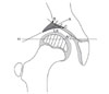
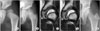
Labral angle (LA)
We define LA as the angle which the labrum makes with the acetabulum (Fig. 1). The labral angle is a direct reflection of the degree of concentric reduction of the hip joint. When the femoral head pushes upward on the labrum (causing high values of AI and CHDD in radiographs), the labrum has a more horizontal position than it would normally. Thus, the labral angle is usually higher in the affected (dysplastic) side than in the unaffected side. The LA normally decreases (that is, the labrum moves downward) in the unaffected side as the hip moves from neutral to abduction and increases as the hip moves from neutral to adduction.
Uncorrected labral deformity (ULD)
In many dysplastic hips the labrum is less elastic than normal, and in abduction the labrum does not return to the normal position (the angle of the labrum on the unaffected side). For that reason, we define the ULD as the difference between the LA of the affected hip in abduction and the LA of the unaffected hip in the neutral position to measure the amount of uncorrected or "residual" deformation of the labrum in the affected hip. If the value of the ULD is close to 0°, the position of the labrum in the affected hip is similar to that of the unaffected hip in neutral. Therefore, the labrum has sufficient elasticity to return to a normal position in abduction.
If the value of ULD is > 0°, the labrum is deformed even when there is no pressure from the femoral head, and more information is needed to evaluate the condition of the hip and the proper treatment.
As a way of confirming the novel information content of the new parameter ULD, we measured the AI and CHDD from radiographs and the ULD from MRI for all of our patients, and defined patient groups based on these parameters, then looked for agreement between the groups thus defined. Little agreement would be interpreted to mean that AI and CHDD do not reflect the condition of the labrum, thus supporting the need for the new parameter (ULD). Radiographs and MRI used for this purpose were taken within 1 week of each other.
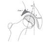
Zone of compressive force (ZCF)
The new parameter, ZCF, is used to represent the amount of lateralization of the femoral head (as does CHDD in radiographs). ZCF (Fig. 2) was determined to evaluate the affected hip in relation to the compressive forces acting on it. According to Pauwells,18) the resultant force of the partial body weight and the abductor muscle group will act in a downward and lateral direction, 16° off the vertical line. In a normal hip, the ZCF is zone 3 in the neutral position and abduction; in a dysplastic hip, the ZCF shifts from zone 2 in the neutral position to zone 3 in abduction, or zone 2 in both positions. The ZCF, along with the change in the LA, facilitated visualization of the degree of reduction of the femoral head.
Calculation of Measurement Parameters
All ROM-MR image data and radiographs were stored in the picture archiving and communication system (PACS; Marosis m-view™ 4.5, Marotech Inc., Seoul, Korea) which provides measurement tools, such as a ruler and goniometer, on the monitor. All parameters were measured 3 times on the 19-inch monitor, then averaged.
Statistical Analysis
The significance of differences in measured parameters between the affected and unaffected hips was analyzed with the Mann-Whitney U-test. The Wilcoxon signed-rank test was used for comparing parameters between hip positions. The agreement between patient groups defined by criteria from radiographs (AI and CHDD) and from ROM-MRI (ULD) was tested in two ways (the simple [unweighted] kappa value and Fisher's exact test). All analyses were performed using SPSS ver. 12.0 (SPSS Inc., Chicago, IL, USA). p-values < 0.05 were considered statistically significant.
RESULTS
Parameters Measured from ROM-MRI
Parameter values in the abduction-internal rotation and abduction-internal rotation-flexion positions did not differ significantly from the parameter values in abduction; thus, the former are omitted from this analysis.
LA and ULD
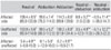
Table 1 gives the values we obtained for LA on the normal and affected sides in the neutral, abduction, and adduction positions when all 25 patients were considered together. In the neutral position, the mean LA of the affected hips was significantly higher than the mean LA of the unaffected hips. Moving from neutral to abduction, the labrum moved downward by a mean of 8.8° and 3.5° in the affected and unaffected hips, respectively. When the hips moved from neutral to adduction, the labrum moved upward by a mean of 5.9° on the affected side, and by a mean of 3.2° on the unaffected side. We believe that the LA changes more on the affected side due to the instability of the joint.
As for ULD, a mean LA of 99.6° in abduction on the affected side was 3.4° smaller than the mean LA of 103.0° of the unaffected side in the neutral position, meaning the mean ULD of our patients was quite favorable. Our 25 affected hips showed the following values of ULD: ULD ≤ 0°, 17 hips; 0° < ULD ≤ 5°, 6 hips; 5° < ULD ≤ 10°, 1 hip; and 10° < ULD ≤ 15°, 1 hip. Thus, 17 (68%) of our affected hips showed "normal" labral elasticity, while 8 (32%) showed residual labral deformity.
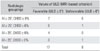
Statistically, the agreement between patient groups defined by AI and ULD was shown to be low, as with CHDD and ULD (Table 2). This was true whether or not we considered each of the 4 radiographic groups separately, as in Table 2, or together (first, by AI value alone, as AI ≥ 25° vs. AI < 25°, or by CHDD value alone, as CHDD ≥ 6% vs. CHDD < 6%). This shows that the radiographic parameters AI and CHDD do not closely reflect (or correspond to) the amount of uncorrected labral deformity.
ZCF
In 10 of the 25 hips (40%), the ZCF shifted from zone 2 in the neutral position to zone 3 in abduction. In the other 15 hips, the ZCF did not change on abduction; the ZCF continued as zone 2 in 8 (32%), and as zone 3 in 7 (28%) hips. In all unaffected hips, the ZCF continued as zone 3 in abduction. Therefore, 17 hips (68%) showed normal reduction of the femoral head into the inner region of the acetabulum in abduction.
As the affected hips moved from the neutral position to adduction, the ZCF shifted from zone 3 to zone 2 in 6 hips and from zone 2 to zone 1 in 2 hips. In the other 17 hips, the ZCF continued as zone 2 in 11 hips and zone 3 in 6. On the normal side, the ZCF continued as zone 3 in 15 hips and zone 2 in 2, while the ZCF shifted from zone 3 to zone 2 in 8. Therefore, in adduction the ZCF was located in the outer region in 19 of the 25 affected hips (76%), but in only 10 of the 25 unaffected hips (40%).
Definition of Concentric Reduction in ROM-MRI
Based on parameters measured from ROM-MRI, we define a concentrically-reduced hip as one in which the affected labrum points downward in the neutral position, at the same angle as the labrum of the normal hip; and in which the ZCF in the neutral position is zone 3 as defined herein (and shown in Fig. 2). For concentric reduction of the femoral head in a dysplastic hip, the LA in hip abduction should be similar to that of the unaffected side in neutral (ULD ≤ 0°) and the medial joint space should be decreased (ZCF moves from zone 2 in neutral to zone 3 in abduction).
DISCUSSION
MR imaging as the hip moves through a range of positions makes it easy to follow the changes in the cartilage and soft tissues, especially the labrum, which has such an important role in the growth of the acetabulum.10-17) As the hip moves from the neutral position to abduction, if the labrum moves downward (that is, if the LA decreases) from a relatively horizontal position, any treatment must reduce the pressure on the labrum (with an abduction brace or a varus-[derotation] osteotomy of the proximal femur), and/or must orient the labrum downward in the normal direction (by a pelvic osteotomy), to facilitate the lateral and downward growth of the lateral part of the acetabulum and its deepening.
Regarding the ULD, our rationale for comparing the LA of the affected hip in abduction with that of the normal hip in the neutral position is that in abduction we see the affected hip when it is freed from pressure and its reduced elasticity is most visible, while on the normal side the position of most importance is the neutral. The ULD is, simply an approximate measure of the amount of correction we would like to achieve by treatment; thus, it does not figure in our definition of concentric reduction.
It could be argued that the ZCF, as here defined, is another measure of lateralization, but a crude one at that. The important point is that the ZCF is a simple guideline obtained from ROM-MR images. The widened joint space visible on radiographs is often confirmed by MRI to be filled with fluid. The femoral head is shifted laterally to a biomechanically disadvantaged position and the ZCF can shift medially in abduction. However, ROM-MRI sometimes reveals that a widened joint space in the radiograph is occupied by a hypertrophied articular cartilage, and is accompanied by a shallow acetabulum with poor head coverage. In such cases, the ZCF does not shift medially in abduction. A V-shaped tear-drop in the radiograph, with a widened superior portion and a thickened acetabular floor, is indicative of residual acetabular dysplasia19) and a reconstructive procedure should be considered.20)
Given the priority we place on rather small changes in the labral angle and the ZCF, we should acknowledge the possibility of intra- or inter-observer error in determining these parameters. Fortunately, the downward or upward shift of the labrum, and the change in acetabular zone, are generally unambiguous and easy to visualize, even without precise measurement or drawing.
It is very difficult to measure the elasticity of the deformed labrum because the biomechanical properties have been changed by the severity and duration of dislocation of the femoral head. Therefore, even after treatment by abduction brace or varus-(derotation) osteotomy of the proximal femur, we cannot predict at what time in the future the ULD will be normalized. However, the return of the labrum to a normal shape and direction is a prerequisite for normal development of the acetabulum, ether by conservative or surgical treatment. A persistently increased LA suggests a high probability of acetabular dysplasia.
Our goal in treating dysplasia is to have a normal hip as early as possible after primary treatment, even before 4-5 years of age,9,20-24) allowing up to 1-2 years following reduction for the acetabular index to return to normal (< 25°). Based on reviews of our patients' radiographs and ROM-MR images, we have created some general guidelines for complete reduction of the femoral head into the acetabulum. By this approach, after the AI and CHDD are confirmed, the next step is to use the ROM-MRI to evaluate the labrum, either by visually comparing the orientation with that of the unaffected side or by measuring the LA and ULD values as we have done. Next, the ZCF would be determined, or the medial joint space would at least be visually inspected. If soon after closed or open reduction plus a hip spica cast the hip is found to be unstable, (i.e., the ULD is ≤ 0° and the ZCF moves from zone 2 in the neutral position to zone 3 in abduction), then the abduction brace treatment should be extended. However, a child of walking age is understandably reluctant to wear a brace. If, 1-2 years after closed or open reduction plus use of an abduction brace the patient shows the same or similar MRI findings, a secondary surgical procedure should be considered (Figs. 3 and 4). If the ULD and ZCF criteria are unfavorable (ULD > 0°, and the ZCF is zone 2 in the neutral position and abduction, meaning that the medial joint space is not decreased in abduction) (Fig. 5), and the medial superior acetabular cartilage is thickened, then both femoral and pelvic osteotomies are required to enhance complete remodeling of the hip. In a case in which the labrum is directed horizontally due to pressure from the femoral head, combined with a hypertrophied articular cartilage, reversal of the pathology is not simply achieved by a brace, or by a unilateral femoral or pelvic osteotomy (Fig. 6).
It is too early to describe our long-term treatment outcomes because osteoarthritis in DDH usually occurs in early or late adulthood, but the results appear to be excellent thus far. Among the patients who underwent surgery, six had another ROM-MRI scan after the metal was removed. In these six patients, the mean LA of the affected hip in the neutral position decreased by 8.7° (from 114.7° to 106.0°) after surgery, and the ZCF was zone 3 (medial) in the neutral position and abduction.
The scanner which we used cannot give a complete picture of the relationship between the diseased femoral head and the acetabulum because it has limited space for the patient's free leg movement. As with arthrography, the images do not show the hip in a weight-bearing position. For younger children who would not tolerate the noise, positioning and being enclosed in the small scanner, the use of sedation is advisable. MR scanning often places a financial burden on the patient's parents. As an inducement for the parents, our institute enables us to offer a discount for a ROM-MRI (3 slices" in each of the 5 positions [T2 only] for one-half of the current cost of a normal standard MR scan).
While we do not wish to disparage the usefulness of the time-honored radiograph for children in this age range, its limitations are quite obvious, and the best use of radiographs will be in conjunction with an imaging technique, such as ROM-MRI, which elucidates the soft tissue structures. ROM-MRI clearly demonstrates the acetabulolabral relationship in detail, which is not possible in radiographs. The convenience of using MRI on an outpatient clinic basis lessens the need for arthrography in the operating room, and the clear images of the labrum and the relationship between the acetabulum and the femoral head enhance accurate decision-making for further treatment.
 XML Download