

 PDF
PDF ePub
ePub Citation
 Print
Print


Ipsilateral fractures of the femur and tibia are called 'floating knee' injuries and may include a combination of diaphyseal, metaphyseal, and intra-articular fractures.1,2) Earlier papers highlighted the elevated risk of complications and permanent disabilities.3,4) Bansal et al.5) and China6) concluded that rigid fixation of both fractures results in excellent or good results. The incidence of fractures resulting from motor vehicle accidents is increasing. Consequently, high velocity accidents are now more common. Such accidents produce violent and complex injuries. Frequently, multiple fractures are produced in the same extremity, adding new dimensions to their management.
These fractures range from simple diaphyseal to complex articular types. This complex injury has increased in proportion to population growth, number of motor vehicles on the road, and high speed traffic. Although the precise incidence of a floating knee is not known, it is a relatively uncommon injury. The largest series reported in the literature was of 222 patients over an 11 year period.7) This injury is generally caused by high-energy trauma with often extensive trauma to the soft tissues. There may also be life-threatening injuries to the head, chest or abdomen and a high incidence of fat embolism. Management of these injuries vary.8) Many of these fractures are open with associated vascular injuries. Surgical stabilization of both fractures and early mobilization of the patient and extremity produce the best clinical outcomes. The use of a radiolucent operating room table and the introduction of retrograde intramedullary fixation of the femoral fractures have facilitated surgical stabilization of some floating-knee fracture patterns. Although treatment planning for each fracture in the extremity should be considered individually to achieve the optimal result, the effect of that decision must be considered in light of the overall injury status of the entire extremity. Collateral ligament and meniscal injuries may also be associated with this fracture complex. Complications, such as compartment syndrome, loss of knee motion, failure to diagnose knee ligament injury, and the need for amputation, are not uncommon. Better results and fewer complications are observed when both fractures are diaphyseal than when one or both are intra-articular.1)
This study examined the outcome of patients after surgical management of a Floating Knee as well as the prognostic factors for this injury.
METHODS
The study included 15 patients (13 men, 2 women; mean age, 34.8 years; range, 18 to 65 years). Children with floating knee injuries were excluded.
According to the classification by Fraser et al.7) the types of the fractures were as follows: type I (5), type IIa (3), IIb (4), and IIc (3). The initial management involved resuscitation and hemodynamic stabilization of the patient, splinting of the affected limb in a Thomas splint and a thorough secondary survey to identify other injuries. Radiographs of the chest, pelvis, affected lower limbs, including all the joints and other suspected bony injuries were performed. Open fractures were observed in two patients, which were classified as Type IIIB according to Gustilo & Anderson's classification.9) Initial wound toilet, tetanus immunization and antibiotic therapy were initiated for open fractures. The floating knee injury was classified according to the Fraser et al.'s classification.7)
Preoperative Evaluation and Initial Management
Patients with floating knee are victims of polytrauma and the involvement of other organs is strongly suspected.
The patients were observed closely for the development of a fat embolism (tachypnoea, confusion, tachycardia). If a fat embolism was diagnosed, the patients were managed in the surgical intensive care and surgical fixation of the fractures was postponed. Patients with associated chest injuries, head injuries or significant abdominal injuries were managed appropriately before surgical stabilization of the fractures.
Once the vital functions were stable, the open fractures were debrided thoroughly in the operating room. The surgical wound was closed only if it was possible without tension. The other wounds always were left open to be closed later, either as a delayed primary closure or with a skin graft.
Femur fractures were treated using locked intramedullary nails in 8 cases (type I: 5 and type IIa: 3), plate-screws in 2 cases (type IIb: 1 and type IIc: 1), and dynamic condylar screws in 5 cases (type IIb: 3 and IIc: 2). Tibia fractures were treated with an external fixator in 2 cases with open fractures, plate-screws in 5 cases (type IIa: 2 and type IIc: 3), or locked intramedullary nailing in 8 cases (type I: 4 and type IIb: 4) (Table 1).
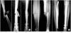
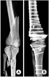
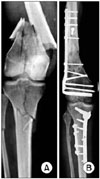
Surgical management of both fractures were performed once the patients were hemodynamically stable and fit to undergo surgery. The femur fracture was fixed prior to the tibia fracture. Intramedullary nailing of both fractures was the commonest method. Both the femur and tibia nails were inserted antegrade. External fixation in open tibia fractures was the definitive management (Figs. 1, 2, 3, 4).
Postoperative Evaluation and Follow-up
Thromboprophylaxis was initiated in all patients during the postoperative period. Physiotherapy was started one week after surgery (isometric for quadriceps & isotonic for hamstring as tolerated). Non weight bearing walking using crutches was permitted for one and half months, followed by partial weight bearing. Full weight bearing was allowed only after clinical and radiological union had been confirmed.
The postoperative clinical, radiological and functional evaluations were performed regularly during the follow-up period. The patients were followed-up every two weeks for one and half months, every month for six months, and every three months thereafter.
The patients were followed up regularly until bony union (clinical and radiological) had been confirmed. A functional assessment and final outcome was measured using the Karlstrom's criteria10) after bony union had been confirmed.
RESULTS
The mean age of the study group was 34.8 years (range, 18 to 65 years). Fourteen patients were involved in motor vehicle accidents whereas one patient sustained the injury from a fall from a height. The right and left sides were involved in 9 and 6 patients, respectively. The average time from admission to surgery was 2 days (range, 1 to 11 days). Intramedullary nailing for both fractures was performed in 4 patients. A combination of a dynamic condylar screw, external fixation for a tibial fracture and buttress plating for tibia plateau fractures were performed in the remaining 9 patients.
The complications encountered were knee stiffness in 2 patients, delayed union of the tibia in 2 patients and superficial infections in one patient.
The additional procedures were manipulation under anesthesia for knee stiffness and dynamization in one patient with delayed union. Patients with delayed union required dynamization of the tibial nail and removal of the external fixator and functional cast bracing of the fracture. These fractures went on to unite after these interventions. The superficial infections were related to the pin sites of the external fixators, which were managed successfully by pin site care and antibiotics. The mean follow-up duration was 23.5 months (range, 21 to 26 months).
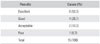
An assessment of the results after bony union according to the Karlstrom criteria revealed the following: excellent, 8; good, 4; acceptable, 2; poor, 1 (Table 2).
DISCUSSION
The term "Floating Knee" is used when the knee joint is isolated partially or completely due to a fracture of the femur and tibia.9) Survivors of high-speed traffic accidents often have injuries to several parenchymal organs as well as multiple fractures. A careful evaluation of these injuries and resuscitation of the patient must precede the definitive management of specific fractures.
Hayes3) suggested that automobile passengers with floating knees should have had their feet braced firmly against the sloping floor of the front seat just prior to the collision, their legs becoming crumpled under the massive decelerating forces produced by the impact. Pedestrians are frequently catapulted some distance from the point of impact and are further injured by striking the pavement. In a study of 222 cases of floating knees by Fraser et al.7) all cases were the result of road traffic accidents.
Studies revealed associated injuries, such as head injuries, chest injuries, abdominal injuries and injuries to the other extremities. Most injuries to the head, chest and abdomen are life threatening. Adamson et al.11) encountered 71% major associated injuries with 21% being vascular injuries. The reported a mortality rate ranging from 5% to 15%, reflecting the seriousness of the associated injuries.1) A deliberate and careful examination of the patient must be carried out to determine is a major intracranial, abdominal or thoracic injury is present. Such injuries should take precedence over extremity injuries in the priority of treatment.
There are different management options for floating knees. Hayes3) suggested that in a patient with multiple fractures in the same extremity, surgical fixation of one or more of the fractures was valuable in the management of the entire limb. Ratliff12) found that internal fixation of the fractures should be done wherever possible as these patients were less likely to develop knee stiffness or shortening and were in hospital and off work for less time than those treated conservatively. Omer et al.4) treated a floating knee using both conservative and surgical fixation, and found that where internal fixation was done for both femoral and tibial fractures, the healing time was approximately 8 weeks earlier than the group managed conservatively. Behr et al.8) treated patients with a floating knee by closed intramedullary nailing with Ender nails and achieved femoral union at an average of 10.3 weeks and tibial union at 18 weeks. Ostrum13) treated patients with a retrograde femoral tibial intramedullary nail through a 4 cm medial parapatellar incision. The mean time to union of the femoral and tibial fractures was 14.7 and 23 weeks, respectively. They opined that this method was an excellent treatment option.
Kao et al.14) summarized the indications of surgical treatment and concluded that patients with closed fractures or open type I or II fractures were treated according to the fracture site. Femoral fractures around the cervicotrochanteric area were fixed under traction with dynamic hip screws, dynamic compression screws, or cannulated screws on a fracture table. Some femoral shaft fractures were fixed with interlocking nails by performing an open reduction with the patient in the decubitus position or by closed reduction under traction on a fracture table. Some femoral shaft fractures were fixed by an open reduction with dynamic compression plates and internal fixation. Femoral fractures in the supra-intercondylar area were fixed by open reduction with condylar plates or dynamic compression screws. Tibial fractures around the tibial plateau, proximal tibia, or distal tibial areas were fixed by open reduction using buttress plates or dynamic compression plates. Some tibial shaft fractures were fixed with interlocking nails by open or closed reduction. Some tibial shaft fractures were fixed by open reduction with dynamic compression plates with internal fixation. For type III open fractures, temporal external skeletal fixators were applied. Internal fixators with plates or nails were applied after the wound condition had stabilized.
Dwyer et al.15) used the combined modalities of treatment with one fracture managed conservatively and the other surgically. They concluded that the treatment method for the tibia did not interfere with joint mobilization.15) Lundy and Johnson1) recommended surgical stabilization of the fractures for early mobilization, which produced the best results. Theodoratus et al.16) recommended intramedullary nailing as the best treatment choice except for grade 3B & C open fractures. The single incision technique for nailing both fractures have been recommended by several authors.7,17,18) Rios et al.18) compared a single incision with traditional antegrade nailing of the fractures and found the former to have less surgical and anesthesia time with reduced blood loss. Shiedts et al.19) reported an increased incidence of fat embolism when both fractures were treated with reamed nails.
Szalay et al.20) demonstrated knee ligament laxity in 53% of patients, whereas 18% complained of instability. Most patients with instability had a rupture of the anterior cruciate ligament with or without damage to the other ligaments. They concluded that a knee ligament injury was more common with floating knee injuries than with isolated femoral fractures, and advocated careful assessment of the knee in all cases of fractures of the femur and floating knee injuries. Other studies21) reported that the incidence of knee ligament injuries in the floating knee was up to 50%, most of which were missed in the initial assessment. A meticulous examination of the knee at the time of injury is strongly advocated, even the practicality of this method is questionable.
The classification used in the present was the one proposed by Fraser et al.,7) which includes intra-articular fractures at the knee.
With this management, the fracture union time and functional recovery was better than the other surgical modalities. This is in accordance with studies reported by Gregory et al.22) and Ostrum,13) who had excellent results with fixation of both fractures by intramedullary nailing. Both authors used retrograde nailing for the femur, even though all nailing was antegrade in the present study. Although no knee problems were found when a single incision technique was used,4,7,17) it is believed that antegrade nailing would allow an easier knee ligament reconstruction if needed because a femoral nail inserted retrograde would make knee ligament reconstruction technically difficult.
Intra-articular involvement of the fractures, higher skeletal injury scores and the severity of soft tissue injuries are significant indicators of a poor outcome.22-24) Hee et al.25) suggested a preoperative scoring system that considered age, smoking status at time of injury, injury severity scores, open fractures, segmental fractures and comminution to affect the prognosis of the final outcome of these fractures.26) The associated injuries played a major role in the initial outcome of patients with regard to a delay in initial surgery, prolonged duration of surgery, anesthetic exposure and a delay in rehabilitation.27)
The best results were obtained when both fractures were treated by intramedullary nailing. These patients returned to their normal level of activity earlier than when the fractures were treated with other modalities. Tibia fractures treated with external fixation had a longer union time, which is probably related to the soft tissue injury and comminution at the initial injury.
The 3 patients who had a poor outcome in the present study were 2 patients with tibia plateau fractures, who had knee stiffness and persisting pain in the knee, and one patient with a grade 3B open tibia fracture treated by external fixation. This shows that the poor prognostic factors are related to the type of fracture (open or closed, intra-articular fractures, severe comminution).
In conclusion, floating knee injuries are a group of complex injuries that require a careful assessment to detect poor prognostic factors (open, intra-articular, comminuted fractures) and associated injuries. Surgical fixation of the fractures with thorough surgical planning and prolonged rehabilitation are recommended. A combination of these determines the ultimate outcome of these patients.

 XML Download