

 PDF
PDF ePub
ePub Citation
 Print
Print


Aseptic loosening of cemented hip prostheses is recognized as a long-term problem, and especially in males and younger patients (Johnsson et al.,1) Callaghan et al.,2) Sochart and Porter3)). Much effort has been focused on developing new prostheses that are designed for cementless fixation. Osseointegration into metal depends on the surface structure and characteristics of the implant.
Central to achieving a lasting clinical result is the ability to achieve immediate and durable stability of the implant to the host bone.4,5) Initial implant stability is achieved at the time of surgery, whereas long-term fixation of a cementless arthroplasty requires osseointegration, which is the firm and reliable adherence of an implant to bone. Because bone is a living tissue, such integration must account for the tendency of bone to remodel over time. Thus, if one expects an implant to exist in situ over the long-term, it must demonstrate not only firm and durable fixation to the bone, but also the absence of adverse bone remodeling or reaction, including stress shielding and osteolysis, which could later compromise the fixation.
The key features of a cementless femoral implant that contributes to its ability to achieve stable bony ingrowth are its design and the surface finish,6) with the former primarily accounting for the initial stability and the latter contributing to the osseous adherence. There is evidence that both features must be favorable for the development of bony stabilization of the implant.6,7) The purpose of this study is to determine the performance of and periprosthetic bone response to a tapered, titanium, hydroxyapatite (HA)-coated hip implant at least 7 years after its insertion.
METHODS
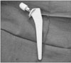
During the years 1998 to 2002, we performed 86 primary total hip replacements in 78 patients. In all cases, we used using a tapered, titanium (Ti6Al4V) HA-coated femoral implant (Corail, Depuy Orthopaedics Inc., Warsaw, IN, USA) (Fig. 1).This femoral implant is available with a collared option, which was used in 22 hips. The remaining 64 hips in this series were inserted without a collar. The titanium tapered implants were entirely coated with a layer of 155 ± 35 µm of HA that was applied with a plasma spray technique. The sizes of the implants used in this series were as follows: 4 size #9, 10 size #10, 19 size #11, 22 size #12, 16 size #13, 6 size #13.5, 6 size #14, 2 size #15, and 1 size #16. The size of the femoral heads used included 36 mm in 20 hips, 32 mm in 50 hips and 28 mm in 16 hips. Although the type of femoral implant was consistent during this experience, a variety of acetabular implants were used, and therefore, their performance was not the target of this investigation.
The average patient age was 59 years (range, 41 to 81 years) among the 35 men (38 hips) and 43 women (48 hips). The diagnoses were primary osteoarthrosis in 56 hips, osteonecrosis in 27 hips, developmental dysplasia of the hip in 1 hip and posttraumatic arthritis in 2 hips. The patients were classified by their activity level and Charnley class,8) with 28 hips being replaced in heavy laborers, 27 in moderate laborers, 14 in light laborers, 14 in semisedentary indivuals and 3 in sedentary individuals. There were 67 hips done in patients who were classified as Charnley class A (1 hip was involved, but there was no other condition that interfered with walking), 8 hips were done in patients who were classified as class B (both hips were involved, but the body was normal and it did not affect walking), and 3 hips were done in patients who were classified as class C (additional factors contributed to the failure of normal walking ability).
Each surgery was performed by one surgeon through a posterolateral approach to the hip with the patient in the lateral position. Patients were allowed to bear weight as tolerated postoperatively and they were given crutches as necessary. All the patients received routine perioperative antibiotics and thromboembolism prophylaxis with subcutaneous heparin.
Clinical evaluation was performed using the scoring system of D'Aubigne and Postel,9) and the hip scores were assigned according to the level of pain, the functional status and the range of motion. Patients who refused to return, but who did forward X-rays for review after being contacted were questioned by phone about the functional status of their hip. All the available radiographs were collected and assessed for implant stability, subsidence, osseointegration, osteolysis, stress shielding and evidence of periprosthetic lucency. The anteroposterior and lateral views of each hip were obtained preoperatively and postoperatively, at six weeks and at three, six and twelve months, and yearly thereafter. The early postoperative views were compared with the films taken at the final follow-up to assess subsidence, acetabular wear and bone remodeling. Subsidence was assessed by measuring any change in the distance from the tip of the greater trochanter to the lateral shoulder of the femoral stem on the sequential radiographs. Varus or valgus positioning and subsidence were determined by comparing the mid axis of the femoral shaft vs. the mid axis of the implant on sequential films. A zonal analysis of the radiolucent lines, as outlined by Gruen et al.,10) and Johnston et al.,11) was used to catalogue the relevant changes in bone morphology and the bone implant interface characteristics. Osseointegration was determined by the presence of spot welds.
RESULTS
As a result, 86 hips (78 patients) were available for review at a follow-up period of more than 7 years. In 11 of the 86 cases, acetabular failure required revision of the acetabular component, but the femoral stem survived and it was available for long-term evaluation. In 2 additional cases, both acetabular and femoral revision were performed due to loosening of the acetabular component. But we also performed femoral component revision for compatibility with the acetabular component. In both cases of femoral revision, the femoral component was well fixed with considerable osseointegration and a femoral osteotomy was required for removal. The acetabular revisions were due to polyethylene wear and osteolysis. In 11 cases the osteolysis was continuous with the joint space. It became encapsulated by a sclerotic margin and it was slowly enlarging. Osteolysis was never a structural problem in the femoral periprosthetic bone. In these cases, there was acetabular loosening and severe acetabular osteolysis. Yet despite this heavy burden of polyethylene particulate debris, osteolysis was never a clinical or radiographic problem on the femoral side.
The radiographs were obtained at 7-year follow-up for another 20 hips, but the patients would not come in for the 7-year clinical evaluation. Therefore, a phone interview was conducted to assess any change in the functional status at a minimum of 7 years.
The patients were questioned in detail about possible hip-related symptoms and changes in the functional status. None of these patients admitted to a problem with their hip and they refused to return for clinical and radiographic documentation because they lived far away and they were not having a problem. None of these patients had had or expected further surgery on their hip. None of the 86 implants that were directly accounted for were revised because of aseptic loosening of the femoral component and there was no evidence of aseptic loosening on any of the follow-up radiographs. The bone remodeling around the implant was generally favorable. There were 4 hips with incomplete radiolucencies, with none affecting more than 30% of the interface.
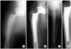
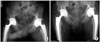
Calcar remodeling was common with rounding of the calcar in 50% of the hips. The group with a collared prosthesis exhibited significantly less calcar rounding. All the femoral implants had evidence that the HA coating had stimulated an osteogenic response by exhibiting evidence of osseointegration with the so-called spot welds or periprosthetic diaphyseal endosteal bone formation seen in at least 3 zones; zones 2 and 3 were the most reliable sites of spot welds (Fig. 2). Subsidence was seen in 7 hips and it was less than 4 mm in each case (Fig. 3). Stress shielding was significant in only 2 cases in which the implants were preferentially bonded distally. The remainder exhibited normal or near-normal preservation of the proximal bone structure.
The clinical scores indicated that the postoperative hip function had improved dramatically when compared with the preoperative levels. The scores improved in the first year and they were maintained throughout the study period unless acetabular failure occurred. The patients requiring acetabular revision had lower scores at the final follow-up, but this was not statistically significant. There was no complaint of thigh pain in any of the patients in this series.
DISCUSSION
This is a study of a prospectively collected series of hip arthroplasties that using a tapered, titanium, HA-coated hip stem. The femoral implant evaluated in this study has been shown to perform quite well,12) and in this group of patients, it developed osseointegration with clear evidence of endosteal spot welds and trabecular bridging in all the cases. There were no cases of aseptic loosening, with a stable bone response reliably demonstrated at greater than 7 years of follow-up. There was no evidence of subsidence or component migration, indicating that implant stability was achieved early and reliably. The results of this study are comparable to those reported by Rokkum and Reigstad13) using this same device. In their review of 94 consecutive cases, with careful radiographic and clinical follow-up, there was no observed stem subsidence or loosening in any case. The authors regularly found cancellous bone remodeling in the proximal femur, with preservation of the osseous architecture. Reikeras and Gunderson14) reported on the performance of this stem at 8 to 12 years of follow-up, and they found only 1 case of mechanical failure among 245 inserted stems. Proximal bone atrophy and distal hypertrophy were infrequent, leading the authors to postulate that the stems had an "essentially physiologic" load profile. Proximal osteolysis was minimal and distal osteolysis was absent, despite a high rate of acetabular failure, and this was all similar to that seen in our cohort. Although the relative contribution of the HA coating to the success of this implant cannot be determined by this study, there is considerable clinical and experimental evidence suggesting that such coatings enhance initial fixation and ultimate osseous integration.5,15) Comparative studies have indicated the advantages of HA-coated implants over the noncoated implants of a similar design,6,16-18) with the HA-coated femoral stems achieving more reliable bony fixation, better evidence of spot welds and less subsidence than the porous control groups. Using dual-energy X-ray absorptiometry, Tanzer et al.18) found that after 2 years of follow-up, the stems with a HA coating exhibited significantly less bone loss than did the stems implanted without a HA coating. In addition, there are a number of reports documenting the long-term success of HA-coated femoral components at greater than 10 years of follow-up19-22) with the absence of fixation failure of the femoral stem seen in 96% to 100% of the cases.
The tapered geometry of this implant may also have played a significant role in the osseous integration and bone adaptation observed in this study. Reliable primary stability for a rapid bone response to HA was probably ensured by the wedge-shaped design of the stem. Long-term success has been documented with other tapered stems without a HA coating.8,23) Although it has been postulated that tapered femoral components optimize the stress transfer from the implant to bone, this current study cannot substantiate that claim. Yet comparing the early postoperative radiographs with the long-term radiographs does demonstrate preservation of the bone's architecture, with a similar appearance of bone quality, both early and late. In addition, the complete absence of thigh pain noted in this group of patients may be related to this favorable bone response that was without significant hypertrophy or remodeling.
Most cementless stems have been coated proximally. The clinical effects of a proximal coating are uncertain. However, with the unacceptable rates of stem revision with proximal porous coating, Engh et al.24) concluded that the fixation of fully coated stems is better than that of stems with a proximal coating. We used an entirely coated stem, and our results are in accordance with the recent long-term results of McNally et al.20) On the other hand, in a multicenter study of 436 proximally HA-coated stems, D'Antonio et al.25) revised 0.46% of the stems because of loosening and they found subsidence of another 6% at a follow-up of 3 years. In another D'Antonio et al.26) study of proximally HA-coated stems, 1% were revised after 2.3 years and 8% experienced subsidence. However, Tonino and Rahmy27) have reported fairly good results with a proximal HA coating. Radiolucency adjacent to a prosthesis has been histologically correlated with a fibrous layer between the bone and the prosthesis by Pidhorz et al.28) We found radiolucent lines adjacent to the stem in 16 cases. In all except 2 cases, this radiolucency was located in the proximal zones. The central and distal zones of the prostheses were otherwise well osseointegrated. Our explanation for the proximal radiolucency is that a well-bonded implant may have some proximal micromovement during loading. Wear particles with a foreign body reaction at the proximal level of the stem may also be of importance. In any case, these observations indicate that an extensive coating may give reliable fixation.
It is noteworthy that despite the high rate of acetabular wear, there were no cases of significant femoral osteolysis. There was no distal osteolysis; when present, lysis was confined by a sclerotic zone next to the proximal zones of the implant and it was not progressive. It is likely that the circumferential, complete coating of the implant conferred upon it a resistance to the progressive and distal manifestations of osteolysis.
In summary, this study documents the success of this tapered, titanium, HA-coated femoral stem in a consecutive series of unselected cases, and there was no failure of fixation or aseptic loosening. The implant was found to perform with equal success across a wide range of pathologic conditions, patient profiles and bone types. Although the relative contributions of the surface coating, the implant geometry and the surgical technique of compaction broaching cannot be established by this study, it is clear that this combination of features resulted in a durable and reliable femoral construct for this group of patients. Further documentation of the performance of this device into the second decade of service and beyond will be necessary to determine if these favorable results are maintained.
 XML Download