

 PDF
PDF ePub
ePub Citation
 Print
Print


Ankylosing spondylitis (AS) is a disease that involves the sacroiliac joint and all the way up to the upper spine. It is characterized by reduced joint motion due to calcification of the spinal ligaments and joint capsules, and it can be accompanied by kyphosis because of changes in either lumbar lordosis or thoracic kyphosis.1,2) Patients with AS experience significant restrictions in their daily activities. In particular, when the disease affects the cervical spine, patients can neither see straight ahead in the standing position nor lie on their back, which disrupts their daily and social activities, necessitating surgical intervention.3,4)
Osteotomy, pedicle screw fixation and bone grafting are popular treatments for kyphosis. The long-term prognostic factors of surgery include the type of osteotomy, the degree of angular correction, and the union of grafted bone. However, there are no reports of nonunion after surgery in AS.5-7) Clinicians are aware through experience that the union of grafted bones is achieved earlier in patients with AS than in those with other spinal diseases. This retrospective study compare the interval from surgical treatment to the union of grafted bone between patients with AS and those with degenerative spinal diseases.
METHODS
Of the AS patients with kyphotic deformity who had been admitted to our institution between January 2003 and January 2009, 10 patients who had been treated with posterolateral fixation after the osteotomy and bone grafting were included in the experimental group. The control group contained 26 patients who had been treated with decompression and posterolateral fusion and bone grafting for degenerative spinal diseases. Nine of the 10 patients with AS underwent pedicle subtraction osteotomy. The remaining patient underwent Smith-Petersen osteotomy. Bone grafting included the two levels adjacent to the osteotomized site. The inclusion criteria for the control group were less than 50 years of age, posterolateral fusion and two-level grafting considering that AS is common in young people and two-level grafting had been performed in the experimental group. In the control group, there were 15, 8, and 3 cases of spinal stenosis, spondylolisthesis, and intervertebral disc herniation, respectively.
Bone grafting was performed with autogenous bone grafts collected during the osteotomy and bone substitutes in the experimental group, whereas it was performed with autogenous bone grafts collected during decompression and autogenous iliac bone grafts in the control group. Bone substitutes were used in 9 cases in the experimental group, (a composite of hydroxyapatite and calcium phosphate in 6, demineralized bone matrix in 2, and an allograft in 1).
Bone union was assessed from the radiographs (anteroposterior, lateral, and oblique views) taken before surgery, after surgery and at regular intervals during the follow-up period. The radiographic results were evaluated using the Lenke's classification system8)
to assess the overall union, and the Christensen's classification system9) to examine the intersegmental union. Bone union was considered to have occurred when type A union (solid bilateral union) and type B union (unilateral large fusion mass with contralateral small fusion mass) according to the Lenke's system or grade 3 union (presence of a bony bridge) according to the Christensen's system were observed on the radiographs. Three-dimensional CT was also performed when bone union was not evident on the radiographs.
The statistical analysis for a comparison of the time to bone union between the two groups was performed using a Mann-Whitney test.
RESULTS
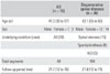
In the experimental group, there were 7 males and 3 females, and the number of fused segments was 40. Their mean age and follow-up period was 41.3 ± 10.7 years (range, 30 to 67 years) and 21.7 months (range, 12 to 43 months), respectively. In the control group, there were 12 males and 14 females, and the number of the fused segments was 104. Their mean age and follow-up period was 43.1 ± 7.4 years (range, 35 to 50 years) and 21.8 months (range, 12 to 74 months), respectively (Table 1). There were no complications, such as infections and nonunion, after surgery in both groups.
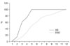
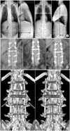
According to the Christensen's classification, grade 3 bone union was achieved in 60% and 100 % of the 40 levels in the experimental group at 3 months and 4 months after surgery, respectively. In the control group, grade 3 bone union was obtained in 50% and 100% of the 104 levels at 5 and 12 months after surgery, respectively (Fig. 1). Bone union could not be assessed using the radiographs alone in 3 cases of the experimental group and 5 cases of the control group. Union could be confirmed with CT in 4 segments in the experimental group and 7 segments in the control group, which were grade 1 or 2 union according to the Christensen's classification system (Fig. 2).
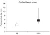
Bone union according to the Lenke's classification system was observed at an average of 3.5 months (range, 3 to 5 months) and 5.6 months (range, 4 to 12 months) after surgery in the experimental group and control group, respectively (Fig. 3), showing a significant difference (p = 0.023).
DISCUSSION
The main pathological features of AS is enthesitis and heterotopic ossification at the insertion of a tendon, ligament or joint capsule into bone causes ankylosis.10) The bone mass is maintained by the balance between the osteoblast and osteoclast activities, and an imbalance in the activities causes heterotopic ossification in AS.
The precise mechanisms of heterotopic ossification and ankylosis need to be established. Bone formation and remodeling are believed to be regulated by Wnt signaling, and the levels of Dickkopf-1 (DKK-1), a Wnt inhibitor, are low in AS patients.11,12) Wnt proteins, which are a family of 19 glycoproteins, have a major impact on osteoblastogenesis and bone growth in fetuses. Signal transduction from the cell-surface receptor to intracellular proteins promotes the differentiation of mesenchymal stem cells into osteoblast precursor cells and osteoblasts followed by bone formation.11-14) DKK-1 and Sfrp1 extracellular proteins are the major intracellular regulators that block Wnt signaling and there are many intracellular regulators. Among them, DKK-1 is believed to be the strongest inhibitor. Diarra et al.15) reported that the inhibition of DKK-1 led to bone formation in the peripheral joints of their animal subjects. Uderhardt et al.16) reported sacroiliac joint ankylosis following a blockade of DKK-1 in a rat model.
Bone morphogenetic proteins (BMP) play a major role in intraarticular heterotopic ossification and ankylosis.17,18) BMP is a member of the TGF-β superfamily, regulates cell proliferation, differentiation and lineage determination, and particularly promotes endochondral bone formation. The levels of BMP-2, BMP-6, and BMP-7 are high in AS patients with BMP-2 involved in early stages and BMP-6 and BMP-7 in the later stages.19)
Rapid bony union in AS patients might be due to the activation of the Wnt pathway and BMP, which promote bone remodeling through bone formation and bone induction.
A range of bone substitutes were used instead of autogenous grafts in the experimental group that are known to be the most effective materials for producing higher union rates than allogenous grafts. Nevertheless, bone union was observed in all AS patients and the interval to bone union was remarkably shorter than in the control group. Radiographs taken 3 months after surgery showed that grade 3 bone union had been achieved in 2, 3, and 4 levels in 7, 2, and 1 case, respectively in the experimental group, whereas it was obtained in 50% of the total levels at 5 months after surgery in the control group.
The limitations of this study are that the study population was small and the cause of early bone union in AS patients was not identified. Therefore, studies involving a large number of subjects focusing on the cause of early union are recommended. Despite these limitations, this study is the first to examine the grafted bone union in AS patients, and the clinical impression of early union in AS patients was confirmed by radiography.
The union of grafted bone was obtained earlier in AS patients treated with an osteotomy than in those with degenerative spinal diseases treated by decompression. Considering the early stage bone loss and high incidence of osteoporosis in AS patients, the early union of grafted bone after surgery for kyphosis is an important factor for determining the time to return to normal life and use a spinal brace.
 XML Download