

 PDF
PDF ePub
ePub Citation
 Print
Print


When performing total hip arthroplasty (THA), sufficient coverage of the acetabular cup by the patient's acetabular bone is crucial for the initial stability of the implant.1-3) In dysplastic hips, such as Developmental dysplasia of the hip sequelae or hip joint infection sequelae, placing the cup in the true acetabulum and acquiring sufficient cup coverage by the subchondral bone may not be possible because of a lack of sufficient host bone. The techniques for managing the problem of insufficient acetabular bone coverage include structural bone graft ing,4-7) the use of a small cup placed in or near the native acetabulum,1) the use of an acetabular reinforcement ring8) and medialization of the acetabular component.2,9,10)
In 1976, Dunn and Hess9) formulated a method that involved intentional medial wall fracture and then cup placement beyond the ilioischial line (the protrusio socket technique) to avoid bone grafting and still achieve cemented acetabular cup stability in dysplastic hips. Hartofilakidis et al.10,11) modified this method by perforating the medial acetabular wall with a reamer instead of an osteotome and they called the technique cotyloplasty. Satisfactory reports were published later concerning the results of implanting cemented cups using cotyloplaty. Dorr et al.2) reported good results when implanting porous-coated acetabular components using this technique.
In this study, we evaluated the results of cotyloplasty performed on dysplastic hips with using a cementless acetabular cup. Our findings demonstrate the need to establish a safety margin for the amount of medial protrusion.
METHODS
From April, 1997 to January, 2003, sixteen dysplastic hips in sixteen patients were treated by cementless THA using the cotyloplasty technique. They were five men and eleven women with a mean age of 47 years (range, 32 to 63 years). In one patient, the operation was performed bilaterally, but cotyloplasty was performed only on the left side. The etiologies of the acetabular dysplasias were developmental in twelve patients and hip joint infection sequelae in four. According to the dysplasia classification of Crowe et al.,12) eight hips were type I, three hips were type II, two hips were type III and three hips were type IV.
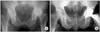
The senior author performed all of the procedures. All the operations were performed with the patient in the lateral position through a posterolateral approach in ten patients and through a lateral approach with trochanteric osteotomy in six patients. Acetabular reaming was done through the medial wall of the true acetabulum until the trial acetabular cup was covered by subchondral bone almost completely, positioned with an inclination of 40 to 45 degrees and with anteversion of 10 to 20 degrees.13) In all cases, the medial wall defect was filled with cancellous bone chips acquired from the resected femoral head and a cementless cup was inserted (Fig. 1). Plasma-sprayed acetabular implants (Plasma cup, Aesculap, Tuttlingen, Germany) were used in fourteen patients, and porouscoated acetabular implants (one Microstructured Omnifit cup and one S-ROM cup) were used in the other two patients. The size of the employed acetabular cup ranged from 44 to 52 and the size of the used femoral head was 28 mm in all the hips. The size of the acetabular cup and the number of screws used in each patient are listed in Table 1.
Postoperatively, the patients were kept in bed for 1 week to 6 weeks, and then they gradually began to mobilize with crutches. The duration of bed rest was subjectively decided on by the operator depending on the amount of cup protrusion and the subjectively assessed intraoperative cup stability. In the early cases, the patients were kept in bed for up to 6 weeks, but this bed rest period decreased as the number of cases increased. Partial weight bearing started at six weeks postoperatively and full weight bearing started at 3 months postoperatively.
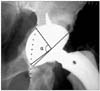
All of the patients had complete clinical and radiographic follow-up. Follow-up clinical and radiographic examinations were performed at six weeks, three months, six months and one year and then annually thereafter. The average follow-up period was 5.9 years (range, 2 to 12.2 years). At each follow-up, a Harris hip score was acquired from the patients' assessments of all items except the range of motion.14) The amounts of hip joint center medialization, cup protrusion beyond the ilioischial line and cup coverage were ascertained on the radiological images. The amount of hip joint center medialization was determined by measuring the horizontal distance between the center of the osseous femoral head preoperatively and the center of the implant femoral head postoperatively on the anteroposterior radiographs of the hip joint.2) The amount of cup protrusion beyond the ilioischial line and the amount of cup coverage were measured as percentages of the 180 degree arc of the cup on the anteroposterior radiographs (Fig. 2).2) Serial radiographs were examined with regard to cup migration, loosening and osteolysis. Loosening of the acetabular cup was defined as a change of the cup position more than 2 mm or a change of the cup angle more than 3 degrees or detection of radiolucent lines around the cup with a line thickness of more than 2 mm.15,16) Periprosthetic cystic or scalloped lesions with a diameter of more than 2 mm that had not been present on the immediate postoperative radiograph were defined as periprosthetic osteolysis.17,18)
RESULTS
The mean preoperative Harris hip score was 57 (range, 24 to 84), and the mean score at the latest follow-up was 94 (range, 89 to 95). Two patients had a mild limp, but these represented improvements over the preoperative limps. One patient had hip joint pain after the operation and another patient had thigh pain, but these symptoms were not associated with loosening and they did not limit the patients' activity.
Stable cup fixation was achieved in all cases except one. At the latest follow-up, two patients showed a radiolucent line narrower than 1 millimeter around the cup; one in zones 1 and 2, and the other in zones 2 and 3.
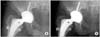
The average amount of hip joint center medialization was 23 mm (range, 9.5 to 51 mm) and the average amount of acetabular cup protrusion beyond the ilioischial line was 44.1% (range, 16 to 66%). The average coverage of the acetabular cup by the host bone was 98.4% (range, 94.4 to 100%). The cancellous bone chips in the medial wall defect consolidated gradually with a reduction in thickness (Fig. 3). The data of the patients is listed in Table 1.
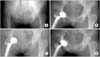
Failure of the initial fixation of the cup occurred in one case. This was the sequela of hip joint infection of a 48-year-old female who showed Crowe type IV dysplasia. In this case, the cup fixation was felt to be stable intraoperatively and the patient started non-weight bearing crutch walking 1 week after operation. However, on the radiographs taken at 2 weeks postoperatively, a change of cup position was detected. The cup then protruded into the pelvis and its inclination decreased from 33.3 degrees immediately after operation to 10 degrees. In this case, the amount of the initial protrusion of the cup beyond the ilioischial line was 66% and its medialization was 51 mm. A revision operation was performed. The cup was easily removed and the medial wall defect was filled with bone graft s harvested from the posterior iliac crest. A new larger size cup (the cup size was changed from 44 to 50) was inserted and this was supplemented with three acetabular screws. This patient was followed for 2.7 years after the revision operation and stable cup fixation was achieved (Fig. 4).
At the latest follow-up, no case showed osteolysis or cup loosening, except for the case of fixation failure mentioned above.
DISCUSSION
Cotyloplasty has advantages over other techniques of fixing an acetabular component in a dysplastic acetabulum. This technique has advantages over superior cup placement because it restores the normal hip joint biomechanics, it restores the leg length discrepancy and it has less chance of impingement that leads to dislocation. It avoids the use of a structural bone graft and it allows hip center medialization. Dorr et al.2) reported that cotyloplasty is a predictable, reproducible method of obtaining proper fixation of a porous-coated acetabular component in a dysplastic acetabulum. In the present study, 2 porous-coated and 14 plasma-sprayed acetabular implants were used, and these plasma-sprayed acetabular implants achieved stable implant fixation. Therefore, cotyloplasty appears to be applicable to various types of cementless acetabular components.
Hartofilakidis et al.10,11) created a controlled fracture of the medial acetabular wall using a reamer. In the present study, instead of fracturing the medial acetabular wall, we perforated the medial acetabular wall by continuous reaming, as was done by Dorr et al.2) who reamed into the quadrilateral plate, and they found that they could prepare a hemispherical osseous cavity more accurately in this manner for the fixation of a metal shell without cement.
After perforating the medial wall of the acetabulum, the defect was filled with cancellous bone chips taken from the resected femoral head, and then an acetabular implant was inserted. Even though a large amount of bone chips was impacted, most of them were gradually resorbed and the remainder became remodeled into a thin cortical bone plate. The thin medial wall might be troublesome when performing revision surgery. Zhang et al.19) introduced a method called acetabular medial wall displacement osteotomy to achieve a thick acetabular medial wall, but this method offers a limited amount of medialization.
For the acetabular reconstructions using a bulk structural bone graft , good results can be expected when less than 50% of the acetabular component is sup ported by the bone graft.4) It is expected that too much medialization through the acetabular medial wall results in initial or late fixation failure. Dorr et al.2) recommended perforating less than 25% of the area of the acetabulum. They reported no fixation failure with up to about 50% of the cup surface beyond the Kohler line. In the present study, initial fixation failure occurred in one case in which the amount of cup surface beyond the Kohler line was 66%. Intraoperatively, the cup was believed to be rigidly fixed, but cup displacement was detected 2 weeks postoperatively. Considering the results of Dorr et al.2) and the results of present study, the safe margin of protrusion in cotyloplasty seems to lie between 50 to 60%. Even through further studies with a larger series of cases are necessary for reaching a more definite conclusion, it seems to be advisable to limit the percent of the protruding cup surface to below 50%. When the coverage is insufficient with 50% of the cup surface beyond the Kohler line, an additional structural bone graft might offer a good solution. In addition, if it is possible, using a larger cup is expected to be recommendable since a smaller cup had less bone contact as compared to a larger cup with the same percentage of protrusion. This also calls for further study.
This study raises a few questions. First, is cotyloplasty needed in type I or II hip dysplasia? Some portion of the patients with type I or II hip dysplasia can be managed with a conventional THA procedure. However, cotyloplasty accompanies medialization of the acetabular cup, which gives an advantage of reducing the joint reaction force and the amount of the load on the acetabulum. This is important for an insufficient acetabulum in a patient with hip dysplasia. Second, what is the specific indication for cotyloplasty as compared to the indications for the other methods, such as using structural bone graft? There is no definite agreement on this matter. This is considered to be more dependent on surgeon's personal choice. The definite indications of each method need further study with a larger number of patients. Yet cotyloplasty is relatively easy to perform and it has demonstrated reliable outcomes, which is a promising advantage.

 XML Download