

 PDF
PDF ePub
ePub Citation
 Print
Print


The term generalized joint laxity (GJL) indicates a generally higher range-of-motion (ROM) than the mean ROM of the general population. The ROM that a joint is capable of is determined by the tightness or otherwise of the restraining ligaments. Joint laxity may be an advantage in sports requiring good flexibility, such as gymnastics. However, it can be potentially dangerous in some other sports.1) Excessive laxity has been associated with a higher likelihood of knee ligament injury2-4) and it is widely accepted that GJL and hyperextension of the knee are important risk factors for an anterior cruciate ligament (ACL) injury, particularly a non-contact injury.5-8) Prior investigations suggest that greater knee laxity and increased GJL are more prevalent in females.6-9) The growth and development and hormonal fluctuations after puberty might contribute to changes in joint laxity and increase the risk of ACL injury risk in females.8) Recently, joint specific laxity, particularly knee hyperextension has been proposed as an important risk factor for ACL injury.7,8,10,11) In addition, the negative effects that altered foot biomechanics have on the ACL have been an area of interest for many researchers,12-15) which adds to the complexity of GJL and its impact on the knee ligaments, particularly the ACL.
Although it is unclear if GJL is related to the outcome of an ACL reconstruction, it has been observed by some surgeons that conservative treatment often fails in patients with GJL, and there is a high risk of a failure in surgical stabilization, possibly because of the biological composition of autograft tissue, as well as the composition of secondary restraints.16,17) Therefore, an ACL reconstruction in those patients should be undertaken with caution. The characteristics of GJL are affected primarily by the inherent connective tissue extensibility that is determined by the composition of connective tissues and the orientation of the various soft tissue structures.18) Since it is determined genetically, an abnormality in the connective tissue composition will be generalized, which needs to be considered when graft selection for a reconstruction is made. Since there is no consensus regarding the ideal/preferred grafts of choice and the rehabilitation protocol, each step beginning from the clinical evaluation to post operative rehabilitation is important for achieving a better clinical outcome. This article reviews the available literature and shares the experience of the senior author in the treatment of an ACL insufficiency in patients with GJL.
CRITERIA FOR ASSESSING GENERALIZED JOINT LAXITY
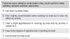
Internationally, there is no agreement regarding the definition of this entity. The criteria for GJL were first described by Carter and Wilkinson in 1964.19) They diagnosed GJL when more than three of the following tests were positive with both upper and lower limbs involved: 1) passive apposition of the thumb to the flexor aspect of the forearm; 2) passive hyperextension of the fingers so that they lie parallel with the extensor aspect of the forearm; 3) ability to hyperextend the elbow more than 10°; 4) ability to hyperextend the knee more than 10°; and 5) an excess range of passive dorsiflexion of the ankle and eversion of the foot. Beighton and Horan20) modified the method described by Carter and Wilkinson in 196919) and revised it in 197321) (Table 1). Of the five joints examined in the Carter and Wilkinson score,19) two were modified. Hyperextension of the fingers to lie parallel to the extensor aspect of the forearm was changed into an ability to perform passive hyperextension of the fifth finger to > 90°, and dorsiflexion of the ankle and eversion of the foot was replaced with a flexion of the trunk. Rotes-Querol22) recommended more tests for the shoulder, cervical spine, hip and toe supplementing the Beighton methods, and different cutoff levels for children and adults. However, many studies have been performed based upon the Beighton methods, even though there is no universal agreement for the GJL criteria between authors using the cutoff level. Clinically, this method has many advantages because it can be carried out very easily without any special measuring instruments, and applies a dichotomous principle. Several studies have reported superior reproducibility and concurrent validity of the Beighton-Horan index than other methods.23-26)
INCIDENCE OF GENERALIZED JOINT LAXITY
Population studies demonstrate wide variations in the prevalence of GJL, which is affected by age, gender and ethnicity.21,27-30) Some authors confirmed that increased GJL was more common in adolescent girls than boys and de creases with age from childhood onward.3,31-34) GJL is ob served more often in Asians and Africans than Caucasians.21,35,36) From the surveys reported, GJL may be present in 2% to 29% of males and 6% to 57% of females.21,37-39) However, most studies focused on young adults. From those that have examined general populations, it would appear that GJL has an overall prevalence of 5% to 20%.30,40) Such a large variation may be explained by the use of different measuring instruments and different cutoff points in the Beighton-Horan index. Several studies have shown a correlation between GJL and occupation. The prevalence of GJL was reported to be significantly higher in ballet dancers than a control group.2,41) American music students and Swedish industrial workers had a relatively high prevalence of GJL.32) Al-Rawi et al.40) reported that the right side (usually dominant side) was significantly less mobile than the left.
GENERALIZED JOINT LAXITY ON RISK OF ANTERIOR CRUCIATE LIGAMENT INJURY
The risk factors that predispose a person to an ACL injury vary. These may be intrinsic non-changeable factors, such as physiological joint laxity, female gender or the size of the femoral notch, and extrinsic, potentially changeable factors, such as the type of footwear, playing surface and inherited conditioning skills and co-ordination.9,42-44) To date, the role that GJL plays in ACL tears is not completely understood.9) After following up 139 professional football players, Nicholas4) reported that football players categorized as having "loose joints" incurred more knee injuries than teammates with "tight joints." In a study reported by Ramesh et al.,8) it was observed that the prevalence of GJL in those who presented for an ACL reconstruction was 42.6% (72 of 169) whereas it was 21.5% (14 of 65) in the control group. Ostenberg and Roos45) registered prospectively injuries in 123 female soccer players and examined their correlation to potential risk factors. They reported that women with GJL had a 5.3 times higher odds ratio of a lower extremity injury than in those without GJL. Similarly, Soderman et al.11) examined prospectively risk factors for leg injuries in 146 female soccer players and reported that the odds ratio of GJL was 3.1. Recently, Uhorchak et al.46) performed a comprehensive study of 859 military academy cadets and reported that subjects with non-contact ACL injuries had significantly more knee laxity and GJL than healthy controls.
Few studies were designed to examine the variables of GJL. In an investigation by Alfred and Bach,17) GJL was assessed in normal controls and ACL-deficient populations to determine if this factor affected the KT-2000 displacements. The thumb-to-forearm laxity (TFL), metacarpophalangeal extension (MPE), elbow recurvatum and knee recurvatum were measured and graded. Based on these observations, MPE and TFL were found to be most important parameters of the four tested. Harner et al.47) in their retrospective study suggested that experimental group was significantly more flexible in the MPE test than the control group. However, in a recent prospective control study, Myer et al.7) reported that measures of knee hyperextension-predicted ACL injury status and a positive measure of knee hyperextension increased the odds of an ACL injury status 5-fold. There is sufficient evidence in the literature to suggest that the final pathway of a non-contact ACL rupture could be hyperextension of the knee.10,11) It was found that events leading to the hyperextension of the knee results in increased anterior translation of the tibia. Therefore, in individuals with pre-existing excessive knee hyperextension, the ACL can hit the intercondylar notch and guillotine itself when the knee is subjected to hyperextension. Moreover, an intricate relationship between proprioception, increased laxity and joint injury have also been reported.8) Loudon et al.48) reported that a person with genu recurvatum has poor proprioceptive control at the terminal degrees of extension. The poor proprioceptive feedback observed in hyperextension and increased joint laxity can affect both limbs and reduce the ability to initiate the protective reflexes.
During weight bearing, the foot and knee act as inter active segments, with pronation of the foot and intern al rotation of the tibia occurring simultaneously. Pro longed pro nation of the foot produces the excessive internal tibial rotation, which may have a preloading effect on the ACL. Thereupon, athletes who abnormally pronate may be more prone to an injury of this ligament.12,13,15,49) Although there is no consensus regarding flat feet and GJL, several articles suggested that a flexible flatfoot was much more common in hypermobile children.50-52)
CLINICAL EVALUATION AND PREOPERATIVE PLANNING
A detailed clinical examination is of utmost importance in patients with an ACL deficient knee and is associated joint laxity. Patients should be evaluated by the clinical tests for an ACL insufficiency and radiological investigations. Radiological studies not only assist in diagnosis but also provide valuable information on associated lesions. Limb alignment should be checked using full length radiographs. Magnetic resonance imaging is a widely used diagnostic tool for knee ligament injuries that also assists in the selection of an autograft for reconstruction.53,54) The universally accepted screening criteria for GJL, the Beighton-Horan index,21) have the limitation that they do not include flatfeet. Moreover, and the potential risk of each variable of the Beighton-Horan index21) is not determined. It is unclear if flatfeet as a criterion for GJL should be included. If a conclusion can be made from future biomechanical and clinical studies analyzing these factors, then a modified criterion should be devised and be followed while evaluating a patient with an ACL deficient knee and GJL.
RECONSTRUCTION OPTIONS AND GRAFT SELECTION
Although there is some concern regarding the inherent laxity of the autograft tissue and laxity of the secondary knee restraints in patients with generalized laxity, an autograft has to be preferred over an allograft for an ACL reconstruction because the latter would be an inferior choice due to its delayed incorporation into bony tunnels of the host and the residual laxity that it produces.55,56) Moreover, there are few reports on ACL reconstructions in a laxity group, and none on the results of an allograft reconstruction. Reports regarding the side to side differences with the widely used semitendinosus-gracilis and bone-patellar tendon grafts are controversial. Some have shown better results with bone patellar tendon grafts,57-59) whereas others have observed comparable results.60-62) There have been some studies on the increased laxity over time observed with semitendinosus-gracilis grafts, particularly in female patients.63,64) A combination of physiologic laxity, a smaller diameter of hamstring tendons and the delayed incorporation of hamstring tendons into the tunnels are some of the reasons for the disappointing results with a soft tissue graft. A recent study also reported inferior results after an ACL reconstruction in patients with excessive ligament laxity, but the results were not significant.6) Patients with excessive knee hyperextension showed inferior results after a reconstruction.65) Laxity of the secondary knee restraints and the increased graft impingement against the intercondylar roof might be the contributing factors for the negative effects of knee hyper extension on a reconstructed graft. There is a paucity of information regarding the relationship between knee hyperextension and stress concentration on the reconstructed graft. It is possible that in patients with knee hyperextension, the reconstructed graft may experience increased stress compared to that on the graft in a knee with normal joint laxity. This may be due to the absence of sufficiently taut ligaments and tendons, which stabilizes the knee and absorbs the ground reaction forces. In addition, there can be increased impingement of the reconstructed graft against the intercondylar roof in knees with excessive hyperextension, which in turn results in graft deterioration or re-rupture. Jagodzinski et al.,66) in a cinematographic magnetic resonance imaging study, reported that impingement between the ACL and the intercondylar roof occurred at 6.3° hyperextension. They emphasized the importance of posterior placement of the tibial tunnel to avoid impingement in knees with increased hyperextension. However, the posterior tibial tunnel can result in an impingement of the graft on the PCL. This was also reported by Nishimori et al.67) With a posteriorly placed tibial tunnel, they reported that 52% (22 out of 42) of reconstructed ACL grafts had impingement on the PCL at the 12 month follow-up. Moreover, posterior placement of the tibial tunnel can result in an elongation of the graft in extension and also vertical tilting of the graft, which might not be ideal for withstanding the anterior draw forces. Therefore, graft selection is critical to overcome these drawbacks in patients with knee hyperextension. Further studies comparing different grafts will be needed to determine the ideal option for this subset of patients.
POSTOPERATIVE REHABILITATION
The importance and merits of accelerated postoperative rehabilitation after an ACL reconstruction has been discussed extensively.68,69) The rehabilitation protocol in our clinic consists of immediate postoperative weight bearing, tolerable, and a full range of motion without protection except for twisting exercises. By three months, strengthening exercises and low force exercises, such as swimming and cycling, can be permitted. Sports activities that involve jumping, pivoting and sidestepping should be allowed only after 6 months. However, whether this accelerated rehabilitation can also be followed in laxity patients is unclear. The healing process is known to vary among patients. Patients with GJL tend to be "slow healers" and may need to be protected longer. Hardin et al.16) suggested that a "decelerated" rehabilitation program might be suitable for this population.
CLINICAL OUTCOME ASSESSMENT
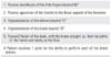
The common methods for evaluating the clinical results can also be used in laxity patients. It should include methods to assess the AP and rotational stability as well as the functional outcome. Tunnels should be evaluated using post operative radiographs. The range of motion can be determined using a goniometer. Anteroposterior laxity can be assessed with a Lachman test and KT-2000 arthrometer. A pivot shift test and the performance of a knee twisting questionnaire (Table 2) can provide information on the rotational stability. The International Knee Documentation Committee and the Lysholm scores can be used for a functional outcome assessment. The reliability of the pivot shift test in assessing the rotational stability has been questioned and recently, the senior author reported that a combination of a pivot shift and the performance of the knee twisting score can be more effective in this regard.54) The mean anterior translation of the uninjured contralateral knee, as measured using a KT-2000 arthrometer, was reported to be higher in patients with GJL than in their the normal counterparts.17) Therefore, the methods used to assess the postoperative stability of knee in these patients should be interpreted with caution.
LESSONS LEARNED OVER THE YEARS
From 2002 to 2005, the senior author treated 72 patients (males, 28; females, 44; age group ranging from 18 to 42 years) with generalized laxity for an ACL insufficiency. Three different autografts (semitendinosusgracilis graft, 11; bone-patellar tendon-bone graft, 32; quadriceps tendon-bone graft, 29) were used for a surgical reconstruction of the ligament in these patients. A semi tendinosus-gracilis graft and bone-patellar tendon-bone graft was used for a single bundle reconstruction and a quadriceps tendon-bone graft was used for a double bundle reconstruction. The selection of a quadriceps tendon-bone graft and the decision for double bundle reconstruction was made based on the thickness of the tendon, as measured by magnetic resonance imaging (selected if the thickness of the tendon was > 7 mm). The choice between a semitendinosusgracilis graft and bone-patellar tendon-bone graft is surgeon dependant. All the patients met the following criteria: 1) they had a unilateral, isolated ligament injury without an injury to the contra-lateral knee; 2) generalized laxity score ≥ 4 points based on the Beighton and Horan index, of which the uninjured contralateral knee showing hyperextension > 10° was considered to be essential; 3) no history of surgery involving the lower extremity, 4) no articular cartilage erosion more than grade II (fissuring and fragmentation < 13 mm in diameter) according to the Outerbridge classification at the time of surgery; 5) no associated ligament injuries of the involved knee; 6) a meniscectomy, when performed, involved less than one-third of the entire meniscus; 7) older than eighteen years; and 8) no axial or rotational malalignment of the involved extremity.
SURGICAL TECHNIQUES
Bone-Patellar Tendon-Bone Autograft
A 10 mm wide bone-patellar tendon-bone graft with a 25 × 8 mm trapezoidal patellar and tibial bone blocks was harvested. The patellar paratenon was sutured. The tibial and femoral tunnels were drilled using a 10 mm diameter cannulated reamer. The femoral tunnel was positioned at the 10:30 o'clock position in the right knee and at the 1:30 o'clock position in the left knee to a depth of 30 mm. After passing the prepared graft through the tibial tunnel, across the joint into the femoral socket, the graft was secured within the femoral socket using an absorbable interference screw. The knee was then moved ten times through the full range of motion, with the graft pulled out in order to pretension it before being fixed within the tibial tunnel with an absorbable interference screw. Femoral socket fixation was performed with the knee in 100° to 110° of flexion while tibial tunnel fixation was done in 10° to 15° of knee flexion.
Semitendinosus-Gracilis Autograft
The hamstring tendons were divided proximally at their musculotendinous junction using an open-loop tendon stripper without detaching their distal insertions.70) The ends of both the gracilis and semitendinosus tendons were whipstiched with #1 Ethibond (Ethicon, Somerville, NJ, USA). A 10 mm diameter reamer was used to drill the tibial tunnel and to create a femoral socket. The femoral socket was reamed to a depth of 30 mm at the position, as described earlier, and then extended using a 4.5 mm diameter reamer as the EndoButton tunnel (Acufex Microsurgical, Andover, MA, USA). Suspensory fixation of the proximal looped tendon was performed in the femoral socket using a looped mersilene tape that was passed through the Endo-Button and secured to the lateral femoral cortex. The leading sutures of both grafts were tied together and pulled through the tibial tunnel with a Kelly clamp to ensure that equal tension was applied to the grafts. The graft was then pretensioned, as mentioned earlier, and when all four strands demonstrated the same amount of tension, buckle staples (Smith and Nephew, Memphis, TN, USA) were used to fix the graft to the tibial cortex distally. Additional fixation of graft in the tibial tunnel and femoral socket were then performed using absorbable interference screws.
Double-Bundle Reconstruction Using Quadriceps Tendon-Bone Autograft
The single tibial tunnel-two femoral socket technique of a double bundle ACL reconstruction described by the senior author was used.53) A full thickness quadriceps tendon including all three tendon layers, 55 mm in length and 11 mm in width, with an attached rectangular shaped patellar bone block (20 mm long, 11 mm wide, 8 mm deep) was harvested. The tendon was then split coronally into two bundles at a ratio of approximately 2:3 and each bundle was then sutured to a length of 30 mm using baseball stitches. Two drill-holes were made in the patellar bone plug through which number-2 Ethibond sutures were passed and Endopearl devices (Linvatec, Largo, FL, USA), 7 mm and 9 mm diameter, were fixed to each tendon end with number-2 Ethibond sutures. A tibial tunnel, 11 mm in diameter, was drilled and the anteromedial femoral socket was created to a depth of 45 mm using the transtibial technique at approximately the 11 o'clock (right knee) or 1 o'clock (left knee) position with a 9 mm cannulated reamer. The femoral socket, 7 mm in diameter, for the posterolateral bundle was created to a depth of approximately 35 mm through an accessory anteromedial portal located 1.5 cm medial to the anteromedial portal immediately above the medial meniscus and 5 mm anterior to the medial femoral condyle. This socket was placed in the centre of the posterolateral bundle footprint, which was located approximately at the crossing point of the long axis line of the ACL attachment and a vertical line drawn through the contact point between the femoral condyle and tibial plateau at 90° of knee flexion. This corresponds to the 9 o'clock and 3 o'clock positions of the right and left knee, respectively. After widening the anteromedial portal to facilitate easy passage of the graft, a leading suture was passed through the tibial tunnel and pulled out through the tibial tunnel. The distal leading sutures attached to the end of the bone plug were pulled out through the tibial tunnel. The leading sutures passed through the corresponding femoral sockets were also pulled out distally through the anteromedial portal. These leading sutures were then tied to the sutures in each limb of the tendon graft and pulled into the respective femoral sockets. The distal bone plug that passed through the anteromedial portal was then placed into the tibial tunnel by pulling the previously passed leading sutures distally. The proximal leading sutures were used to guide the grafts into the femoral sockets. Both femoral and tibial fixation was achieved using bioabsorbable interference screws. The femoral fixation of a posterolateral bundle graft was performed with the knee at 90° of flexion and the screw inserted through the low anteromedial portal. The graft was then pre-tensioned, as described earlier, and tibial fixation of the distal bone plug was performed with the knee in 10° flexion. Finally, anteromedial bundle graft fixation was achieved through the low anteromedial portal with the knee in 70° flexion. Some problems with this technique, such as a difficulty in passing the graft, the need to remove the ACL remnant and widen the anteromedial portal to facilitate graft passage, have been pointed out, and recently a revised technique was described to overcome them.71) This modified, remnant preserving technique has three distinctive features in terms of the rotation- direction of the reamer, graft preparation and passage.
Outcomes and Clinical Evaluation of Each Graft
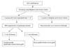
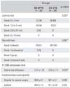
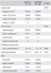
When the clinical outcomes of different methods after 2 years of reconstruction were analyzed, the double-bundle quadriceps tendon-bone graft showed better clinical results than single bundle bone-patellar tendon-bone graft in patients with GJL (Table 3).5) When single bundle grafts were compared, the bone-patellar tendon-bone graft and hamstring graft, the soft tissue graft showed inferior results (Table 4).6) Although the clinical results of these grafts are comparable in normal individuals,60-62) graft site morbidity with a bone tendon graft has resulted in wide acceptance of a soft tissue graft. However, in patients with excessive laxity, bone tendon grafts scores higher than soft tissue grafts even when graft site morbidity is considered. Therefore from the observations made on the 72 patients studied, it is reasonable to favor a double-bundle quadriceps tendon-bone graft for ACL reconstruction over the other two autografts. When a single bundle reconstruction has to be undertaken, the bone-patella tendon-bone graft is preferred. The algorithm (Fig. 1) shows the protocol that can be followed while treating an ACL insufficiency in patients with GJL.
CONCLUSION
The observations of the senior author and an analysis of the relevant literature highlight the challenges of an ACL reconstruction in patients with GJL. The complexity of this entity shows that special attention to all the contributing factors is necessary to achieve satisfactory results in this group of patients. A careful preoperative evaluation, proper surgical technique and well monitored post operative rehabilitation, affect the final outcome after a ligament reconstruction.
 XML Download