

 PDF
PDF ePub
ePub Citation
 Print
Print


Woo-Kyung Son, Seung-Yun Shin, Seung-Min Yang , Seung-Beom Kye
, Seung-Beom Kye
Abstract
Purpose
The aim of this report is to investigate the efficacy of anorganic bovine bone xenograft(Bio-Oss®) at maxillary sinus floor augmentation.
Materials and methods
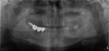
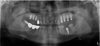
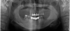
Two male patients who missed maxillary posterior teeth were included. They were performed maxillary sinus floor augmentation using anorganic bovine bone xenograft(Bio-Oss®). After 10 or 13 months, the regenerated tissues were harvested using trephine drills with 2 or 4mm diameter and non-decalcified specimens were made. The specimens were examined histologically and histomorphometrically to investigate graft resorption and new bone formation.
Results
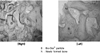
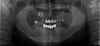
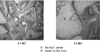
Newly formed bone was in contact with Bio-Oss® particles directly without any gap between the bone and the particles. The proportions of newly formed bone were 23.4~25.3% in patient 1(Pt.1) and 28.8% in patient 2(Pt.2). And the proportions of remained Bio-Oss® were 29.7~30.2% in Pt.1 and 29.2% in Pt.2. The fixtures installed at augmented area showed good stability and the augmented bone height was maintained well.
Figures and Tables


References
1. Bays R. The pathophysiology and anatomy of edentulous bone loss. Reconstructive Preprosthetic Oral and Maxillofacial Surgery. 1986. 1:1–17.
2. Galindo-Moreno P, Avila G, Fernandez-Barbero JE, et al. Evaluation of sinus floor elevation using a composite bone graft mixture. Clin Oral Impl Res. 2007. 18:376–382.

3. Zinner ID, Small SA. Maxillary sinus grafts and prosthetic management. Implant Dentistry:From Failure to Success. 2004. 1st edition. Hong kong: Quintessence Books;99–100.
4. Kamada M, Shimazu K, Aoki H, et al. Maxillary sinusitis caused by oral implants. Practica Oto-Rhino-Laryngologic. 2003. 96:231–236.
5. Galindo-Moreno P, Sanchez-Fernandez E, Avila G, et al. Migration of implants into the maxillary sinus: two clinical cases. Int J Oral Maxillofac Implants. 2005. 20:291–295.
6. Van den Bergh JPA, Ten Bruggenkate CM, Disch FJM, et al. Anatomical aspects of sinus floor elevation. Clin Oral Impl Res. 2000. 11:256–265.
7. Jensen OT. The Sinus Bone Graft. 2006. 2nd edition. Quintessence Books;211–219.
8. Davies JE. In vitro modeling of the bone/implant interface. Anatomical Record. 1996. 245:426–445.
9. Wallace SS, Froum SJ. Effect of Maxillary sinus augmentation on the survival of endosseous dental implants. A systematic review. Ann Periodontol. 2003. 8:328–343.
10. Del Fabbro M, Testori T, Francetti L, et al. Systematic review of survival rates for implants placed in the grafted maxillary sinus. Int J Periodontics Restorative Dent. 2004. 24:565–577.
11. Daelemans P, Hermans M, Godet F, et al. Autologous bone graft to augment the maxillary sinus in conjunction with immediate endosseous implants: a retrospective study up to 5 years. Int J Periodontics Restorative Dent. 1997. 17:27–39.
12. Hallman M, Sennerby L, Lundgren S. A clinical and histologic evaluation of implant integration in the posterior maxilla after sinus floor augmentation with autogenous bone, bovine hydroxyapatite, or a 20:80 mixture. Int J Oral Maxillofac Implants. 2002. 17:635–643.
13. Valentini P, Abensur DJ. Maxillary sinus grafting with anorganic bovine bone: A clinical report of long-term results. Int J Oral Maxillofac Implants. 2003. 18:556–560.
14. Piattelli M, Favero GA, Scarano A, et al. Bone reactions to anorganic bovine bone(BioOss) used in sinus augmentation procedures: A histologic long-term report of 20 cases in humans. Int J Oral Maxillofac Implants. 1999. 14:835–840.
15. Lee YM, Shin SY, Kim JY, et al. Bone reaction to bovine hydroxyapatite for maxillary sinus floor augmentation: Histologic results in humans. Int J Periodontics Restorative Dent. 2006. 26:471–481.
16. Valentini P, Abensur D, Wenz B, et al. Sinus grafting with porous bone mineral(BioOss) for implant placement: A 5-year study on 15 patients. Int J Periodontics Restorative Dent. 2000. 20:245–253.
17. Hammerle CHF, Chiantella GC, Karring T, et al. The effect of a deproteinized bovine bone mineral on bone regeneration around titanium dental implants. Clin Oral Impl Res. 1998. 9:151–162.
18. Valentini P, Abensur D, Densari D, et al. Histological evaluation of BioOss in a 2-stage sinus floor elevation and implantation procedure. A human case report. Clin Oral Impl Res. 1998. 9:59–64.
19. Scarano A, Pecora G, Piattelli M, et al. Osseointegration in a sinus augmented with bovine porous mineral: Histological results in an implant retrieved 4 years after insertion. A case report. J Periodontol. 2004. 75:1161–1166.
 XML Download