

 PDF
PDF ePub
ePub Citation
 Print
Print


Seung-Yun Shin , Seung-Min Yang, Seung-Beom Kye
, Seung-Min Yang, Seung-Beom Kye
Abstract
Purpose
Many researches showed loss of alveolar bone in fresh extraction socket and even in case of immediate implant placement. The aim of this study was to evaluate the effect of non-resorbable barrier membrane on the change of buccal and lingual alveolar bone in immediate implant placement into periapically infected extraction sockets.
Materials and methods
Immediate implants were placed into artificially induced periapical lesion of mandibular premolars after complete debridement using buccal bone defect made by a 6mm trephine bur in 4 mongrel dogs. Before flap repositioning, a non-resorbable barrier membrane was placed on the buccal defect in the experimental group. No membrane was placed in the control group. In 12 weeks after placement, the dogs were sacrificed and undecalcified histologic specimens were prepared. The vertical distance from the smooth-rough surface interface(SRI) to gingiva, 1st bone contact and bone crest were measured in buccal and lingual side. The horizontal thicknesses of gingiva and bone at 0, 1, 2 and 3 mm below SRI were measured.
Results
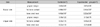
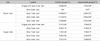
The buccal bone was resorbed more than lingual bone in both groups and there was statistical significance(p<0.05). The distances from SRI to 1st bone contact were 2.45±2.35 mm in experimental group and 4.49±3.10 mm in control group. In all vertical level, lingual bone was thicker than buccal bone(p<0.05).
Figures and Tables
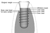 | Figure 1Schematic drawing presenting the location of various histometric measurements(SRI, smooth-rough surface interface; 0mm level, the vertical point for measuring the horizontal thickness of gingiva and bone; 1mm, 2 mm and 3mm level, the vertical point for measuring the horizontal thickness of bone).
|
References
1. Cardaropoli G, Araujo M, Lindhe J. Dynamics of bone tissue formation in tooth extraction sites. An experimental study in dogs. J Clin Periodontol. 2003. 30:809–818.
2. Carlsson GE, Thilander H, Hedegard B. Histologic changes in the upper alveolar process after extractions with or without insertion of an immediate full denture. Acta Odontol Scand. 1967. 25:21–43.

3. Bahat O, Deeb C, Golden T, Komarnyckij O. Preservation of ridges utilizing hydroxyapatite. Int J Periodontics Restorative Dent. 1987. 7:34–41.
4. Park J, Kwon Y, Park J, et al. The effect of human demineralized freeze-dried xenograft on vertical bone formation in beagle dogs. J Korean Acad Periodontol. 2008. 38:75–82.
5. Seibert JS, Salama H. Alveolar ridge preservation and reconstruction. Periodontol 2000. 1996. 11:69–84.
6. Paolantonio M, Dolci M, Scarano A, et al. Immediate implantation in fresh extraction sockets. A controlled clinical and histological study in man. J Periodontol. 2001. 72:1560–1571.
7. Araujo MG, Sukekava F, Wennstrom JL, Lindhe J. Ridge alterations following implant placement in fresh extraction sockets: an experimental study in the dog. J Clin Periodontol. 2005. 32:645–652.
8. Botticelli D, Berglundh T, Lindhe J. Hard-tissue alterations following immediate implant placement in extraction sites. J Clin Periodontol. 2004. 31:820–828.
9. Chang SW, Shin SY, Hong JR, et al. Immediate implant placement into infected and noninfected extraction sockets: a pilot study. Oral Surg Oral Med Oral Pathol Oral Radiol Endod. 2009. 107:197–203.
10. Nevins M, Camelo M, De Paoli S, et al. A study of the fate of the buccal wall of extraction sockets of teeth with prominent roots. Int J Periodontics Restorative Dent. 2006. 26:19–29.
11. Fickl S, Zuhr O, Wachtel H, Bolz W, Huerzeler MB. Hard tissue alterations after socket preservation: an experimental study in the beagle dog. Clin Oral Implants Res. 2008. 19:1111–1118.
12. Antoun H, Sitbon JM, Martinez H, Missika P. A prospective randomized study comparing two techniques of bone augmentation: onlay graft alone or associated with a membrane. Clin Oral Implants Res. 2001. 12:632–639.
13. Lorenzoni M, Pertl C, Polansky RA, Jakse N, Wegscheider WA. Evaluation of implants placed with barrier membranes. A restrospective follow-up study up to five years. Clin Oral Implants Res. 2002. 13:274–280.
 XML Download