

 PDF
PDF Citation
 Print
Print


Abstract
Purpose
The purpose of this report is to show three cases treated by an intergrated periodontal and restorative dentistry approach.
Methods
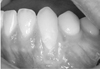
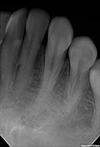
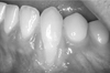
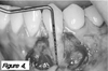
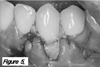
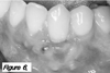
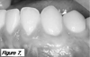
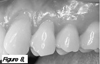
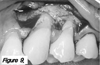
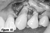
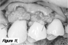
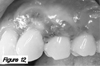
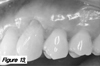
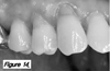
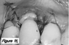
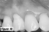
Three patients with Miller Class I gingiva recessions associated with cervical lesions were enrolled for treatment. Two patients received a connective tissue graft and resin modified glass ionomer, and one patient was treated with a connective tissue graft, resin restoration. Keratinized gingiva and relative gingival recession were measured.
Results
The mean reduction of relative gingival recession was 3.7 mm, and the mean keratinized gingiva increase was 2.5 mm. The percentage of root coverage was 80% in average. No signs of gingival inflammation or bleeding on probing were seen. The patients were satisfied with the final esthetics and had no more dentin hypersensitivity.
Conclusions
This report indicates that teeth with Miller Class I gingival recession associated with cervical lesions can be successfully treated by a connective tissue graft combined with restorative dentistry. However, longitudinal randomized controlled clinical trials must be performed to support this approach.
Figures and Tables

References
1. Wennström JL. Mucogingival therapy. Ann Periodontol. 1996. 1:671–701.

2. Löe H, Anerud A, Boysen H. The natural history of periodontal disease in man: Prevalence, severity, and extent of gingival recessions. J Periodontol. 1992. 63:489–495.
3. Serino G, Wennström JL, Lindhe J, Enertoh L. The prevalence and distribution of gingival recession in subjects with high standard of oral hygiene. J Clin Periodontol. 1994. 21:57–63.
4. Sangnes G, Gjermo P. Prevalence of oral soft and hard tissue lesions related to mechanical toothcleansing procedures. Community Dent Oral Epidemiol. 1976. 4:77–83.
5. Toffenetti F, Vanini L, Tammaro S. Gingival recessions and noncarious cervical lesions: A soft and hard tissue challenge. J Esthet Dent. 1998. 10:208–220.
6. Zucchelli G, Testori T, De Sanctis M. Clinical and anatomical factors limiting treatment outcomes of gingival recession: A new method to predetermine the line root coverage. J Periodontol. 2006. 77:714–721.
7. Matis BA, Cochran MA. Technique on restoring cervical lesions. Oper Dent. 2002. 27:525–527.
8. Chan DC, Adkins J. Technique on restoring subgingival cervical lesion. Oper Dent. 2003. 29:350–353.
9. Terry DA, Mcguire MK, Mclaren E, Fulton R, Swift EJ Jr. Perioesthetic approach to the diagnosis and treatment of carious and noncarious cervical lesions: Part II. J Esthet Restor Dent. 2003. 15:284–296.
10. Raetzke PB. Covering localized areas of root exposure employing the "envelope" technique. J Periodontol. 1985. 56:397–402.
11. Langer B, Langer L. Subepithelial connective tissue graft technique for root coverage. J Periodontol. 1985. 56:715–720.
12. Nelson SW. The subepithelial connective tissue graft. A bilaminar reconstructive procedure for the coverage of denuded root surfaces. J Periodontol. 1987. 58:95–102.
13. Dragoo MR. Resin-ionomer and hybrid-ionomer cements: Part II, human clinical and histologic wound healing responses in specific periodontal lesions. Int J Periodontic Restorative Dent. 1997. 17:75–87.
14. Scherer W, Dragoo MR. New subgingival restorative procedures with Geristore resin ionomer. Pract Periodontics Aesthet Dent. 1995. 7:1–4.
15. White C Jr. Repair of a root resorption lesion, A case report. J Periodontol. 1998. 69:596–600.
16. Anderegg CR. The treatment of class III maxillary furcations using a resin-ionomer: A case report. J Periodontol. 1998. 69:948–950.
17. Bruno JF, Bowers GM. Histology of a human biopsy section following the placement of a subepithelial connective tissue graft. Int J Periodontics Restorative Dent. 2000. 20:225–231.
18. Sasanaluckit P, Albustany KR, Doherty PJ, Williams DF. Biocompatibility of glass ionomer cements. Biomaterials. 1993. 14:906–916.
19. el Mallakh BF, Sarkar NK. Fluride release from glass-ionomer cements in de-ionized water and artificial saliva. Dent Mater. 1990. 6:118–122.
20. Breault LG, Fowler EB, Primack PD. Endodontic perforation repair with resin-ionomer: A case report. J Contemp Dent Pract. 2000. 1:48–59.
21. Alkan A, Keskiner I, Yuzbasioglu E. Connective tissue grafting on resin ionomer in localized gingival recession. J Periodontol. 2006. 77:1446–1451.
 XML Download