

 PDF
PDF Citation
 Print
Print


Hyun-Wook Nam1, Yoon-Jeong Park2, Ki-Tae Koo1, Tae-Il Kim1 , Yang-Jo Seol1, Yong-Moo Lee1, Young Gu1, In-Chul Rhyu1, Chong-Pyoung Chung1
, Yang-Jo Seol1, Yong-Moo Lee1, Young Gu1, In-Chul Rhyu1, Chong-Pyoung Chung1
Abstract
Purpose
Following tooth extraction caused by severe periodontitis, alveolar ridge dimension lose their original volume. To reduce the alveolar ridge dimension, the ridge preservation technique has been introduced and tested in many clinical studies with membrane alone or membrane plus graft, achieving reduced ridge loss compared to extraction only. The aim of the present clinical study was to compare the post-extraction dimensional changes in the membrane exposure group to non-exposure group during healing period following ridge preservation technique.
Methods
Ridge preservation was performed in 44 extraction sites. After extraction, deproteinized bovine bone mineral coated with synthetic oligopeptide (Ossgen-X15®) or deproteinized bovine bone mineral (Bio-Oss®) was implanted into the socket. A collagen membrane (Bio-Gide®) was trimmed to cover the socket completely and applied to the entrance of the socket. Four clinical parameters were compared between baseline and 6 months.
Results
During healing period, membrane exposure was observed at 19 sites. At the re-entry, hard newly formed tissue were observed at the ridge preservation site. The grafted socket sites were well preserved in their volume dimension. In both groups, horizontal ridge width was reduced and vertical height was increased. There were not statistically significant differences in horizontal (-1.32 mm vs -1.00 mm) and vertical ridge change (2.24 mm vs 2.37 mm at buccal crest, 1.36 mm vs. 1.53 mm at lingual crest) between two groups.
Figures and Tables
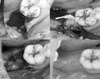 | Figure 1Ridge preservation procedure. (A) Post-extraction view (B) Bone particles and collagen membrane (C) Primary closure (D) 6 months healing view.
|
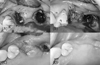 | Figure 2Membrane exposure case (A) After extraction of #25, 27 (B) Ridge preservation on #25 extraction socket (C) 2 weeks of healing after ridge preservation. Membrane was exposed (D) 2 months of healing. Exposed membrane was not observed.
|
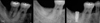 | Figure 3(A) Before extraction (B) 6 months of healing after ridge preservation (C) Implant fixture installation.
|


References
1. Amler MH. The time sequence of tissue regeneration in human extraction wounds. Oral Surg Oral Med Oral Pathol. 1969; 27:309–318.

2. Araujo MG, Lindhe J. Dimensional ridge alterations following tooth extraction. An experimental study in the dog. J Clin Periodontol. 2005; 32:212–218.
3. Schropp L, Wenzel A, Kostopoulos L, Karring T. Bone healing and soft tissue contour changes following single-tooth extraction: a clinical and radiographic 12-month prospective study. Int J Periodontics Restorative Dent. 2003; 23:313–323.
4. Dies F, Etienne D, Abboud NB, Ouhayoun JP. Bone regeneration in extraction sites after immediate placement of an e-PTFE membrane with or without a biomaterial. A report on 12 consecutive cases. Clin Oral Implants Res. 1996; 7:277–285.
5. Becker W, Clokie C, Sennerby L, Urist MR, Becker BE. Histologic findings after implantation and evaluation of different grafting materials and titanium micro screws into extraction sockets: case reports. J Periodontol. 1998; 69:414–421.
6. Artzi Z, Tal H, Dayan D. Porous bovine bone mineral in healing of human extraction sockets. Part 1: histomorphometric evaluations at 9 months. J Periodontol. 2000; 71:1015–1023.
7. Froum S, Cho SC, Rosenberg E, Rohrer M, Tarnow D. Histological comparison of healing extraction sockets implanted with bioactive glass or demineralized freeze-dried bone allograft: a pilot study. J Periodontol. 2002; 73:94–102.
8. Carmagnola D, Adriaens P, Berglundh T. Healing of human extraction sockets filled with Bio-Oss. Clin Oral Implants Res. 2003; 14:137–143.
9. Iasella JM, Greenwell H, Miller RL, et al. Ridge preservation with freeze-dried bone allograft and a collagen membrane compared to extraction alone for implant site development: a clinical and histologic study in humans. J Periodontol. 2003; 74:990–999.
10. Guarnieri R, Pecora G, Fini M, et al. Medical grade calcium sulfate hemihydrate in healing of human extraction sockets: clinical and histological observations at 3 months. J Periodontol. 2004; 75:902–908.
11. Nevins M, Camelo M, De Paoli S, et al. A study of the fate of the buccal wall of extraction sockets of teeth with prominent roots. Int J Periodontics Restorative Dent. 2006; 26:19–29.
12. Pinho MN, Roriz VL, Novaes AB Jr, et al. Titanium membranes in prevention of alveolar collapse after tooth extraction. Implant Dent. 2006; 15:53–61.
13. Barone A, Aldini NN, Fini M, Giardino R, Calvo Guirado JL, Covani U. Xenograft versus extraction alone for ridge preservation after tooth removal: a clinical and histomorphometric study. J Periodontol. 2008; 79:1370–1377.
14. Araujo M, Linder E, Wennstrom J, Lindhe J. The influence of Bio-Oss Collagen on healing of an extraction socket: an experimental study in the dog. Int J Periodontics Restorative Dent. 2008; 28:123–135.
15. Araujo M, Linder E, Lindhe J. Effect of a xenograft on early bone formation in extraction sockets: an experimental study in dog. Clin Oral Implants Res. 2009; 20:1–6.
16. Araujo MG, Lindhe J. Ridge preservation with the use of Bio-Oss collagen: A 6-month study in the dog. Clin Oral Implants Res. 2009; 20:433–440.
17. Norton MR, Odell EW, Thompson ID, Cook RJ. Efficacy of bovine bone mineral for alveolar augmentation: a human histologic study. Clin Oral Implants Res. 2003; 14:775–783.
18. Vasilic N, Henderson R, Jorgenson T, Sutherland E, Carson R. The use of bovine porous bone mineral in combination with collagen membrane or autologous fibrinogen/fibronectin system for ridge preservation following tooth extraction. J Okla Dent Assoc. 2003; 93:33–38.
19. Vance GS, Greenwell H, Miller RL, Hill M, Johnston H, Scheetz JP. Comparison of an allograft in an experimental putty carrier and a bovine-derived xenograft used in ridge preservation: a clinical and histologic study in humans. Int J Oral Maxillofac Implants. 2004; 19:491–497.
20. Simon BI, Von Hagen S, Deasy MJ, Faldu M, Resnansky D. Changes in alveolar bone height and width following ridge augmentation using bone graft and membranes. J Periodontol. 2000; 71:1774–1791.
21. Fickl S, Schneider D, Zuhr O, Hinze M, Ender A, Jung RE, Hürzeler MB. Dimensional changes of the ridge contour after socket preservation and buccal overbuilding: an animal study. Journal of Clinical Periodontology. 2009; 36:442–448.
22. Skoglund A, Hising P, Young C. A clinical and histologic examination in humans of the osseous response to implanted natural bone mineral. Int J Oral Maxillofac Implants. 1997; 12:194–199.
23. Clergeau LP, Danan M, Clergeau-Guerithault S, Brion M. Healing response to anorganic bone implantation in periodontal intrabony defects in dogs Part I Bone regeneration. A microradiographic study. J Periodontol. 1996; 67:140–149.
 XML Download