

 PDF
PDF ePub
ePub Citation
 Print
Print


INTRODUCTION
Periodontal disease is a mixed contamination by periodontal microbes. The activity of the disease is dependent on the susceptibility of the host, an increase in the number of pathogenic microbes and a decrease in the number of advantageous microbes1). Periodontal tissue destruction occurs when the balance of the host's local and systemic immunologic defense system is broken by pathogenic microbes. Those microbes may inhabit not only the oral mucosa, periodontal pocket, tongue, saliva but also the oropharynx and paranasal sinuses. Changing the host's susceptibility is difficult because it is a genetic factor. Therefore, successful periodontal treatment should aim to decrease the number of pathogenic microbes in intra-oral ecological niches and plaque and re-establish an oral environment suitable for normal flora2).
Scaling and root planing (SRP), is a frequently suggested treatment for periodontal contamination3). Most periodontal diseases respond well to nonsurgical treatment but the clinical results are dependent on patient's cooperation, the composition of plaque, and genetic or environmental factors. After mechanical instrumentation, the number of subgingival microbes decreases by one thousandth4). However, in the case of conventional SRP (cSRP), which is performed by quadrants or sextants, pathogenic microbes from untreated areas such as the periodontal pocket, tongue, mucosa, pharynx or saliva can re-cluster into the treated pocket within a week leading to a recurrence of the disease5).
In order to reduce the probability of cross contamination from untreated pockets to treated ones, Quirynen et al. introduced a method for completing the entire SRP under local anesthesia with chlorhexidine as a mouth wash in two-visits within 24 hours. They called it full-mouth disinfection6). The method consists of 1-minute brushing of the dorsal surface of the tongue with a chlorhexidine gel (1%), 1-minute mouth irrigation with a chlorhexidine solution (0.2%) with pharynx in contact with the solution for the last 10 seconds of the rinse. After the completion of each SRP, all pockets should be irrigated subgingivally with a chlorhexidine gel (1%) three times within 10 minutes with the irrigation being repeated after one week. The patient is instructed to mouth-wash with 10ml of a chlorhexidine solution (0.2%) twice daily for 1 minute over a 2-week period.
Many studies have reported the clinical and microbiological effects of full-mouth disinfection (Fdis). After 2 months of Fdis, chronic periodontitis patients showed a significant decrease in the sulcus bleeding index and plaque index. After 8 months, clinical improvements are particularly noticeable in the deep pockets7). Bollen et al. reported that the clinical and microbiological improvements lasted for 4 months8).
Mongardini et al.9) and Cho et al.10) reported clinically improved results after Fdis for aggressive periodontitis patients. However, there are some studies showing that Fdis has no added benefit compared with conventional methods. One study assumed the host's increased immune reaction was caused by bacteremia after Fdis but reported a similar immune reaction to those of cSRP11). Another study reported similar clinical effect to those of cSRP after completion of Fdis in a single visit12-14). A more recent study by Moreira and Feres-Filho reported similar clinical results to those of cSRP after Fdis to aggressive periodontitis patients15). Therefore, the clinical effects of Fdis are not conclusive and there may be differences in the protocol of Fdis and subjects.
In Korea, patients can receive medical insurance benefits only when they received scaling in advance before subgingival SRP, and only a chlorhexidine solution (0.1%) can be purchased over-the-counter. Therefore, the protocol reported by Quirynen et al.6) was modified for clinically easy application to fit the actual situation in Korea.
This study compared the clinical effects of modified Fdis after scaling using a chlorhexidine solution (0.1%) with those of conventional SRP in the treatment of moderate to severe generalized chronic periodontitis for 6 months.
MATERIALS AND METHODS
1. Study Population
Among the patients at the department of periodontology, Chonnam National University Hospital, those who satisfied the conditions below were included in the experiment.
Generalized chronic periodontitis patients with an average bone loss at the time of the first visit between 20~40%, and an attachment loss of more than 3 mm taking more than 30% in the dental arches7). No record of tooth extraction in the upper right arch other than the third molar.
Generally healthy patients (those with systemic diseases, pregnancy and nursing were excluded.)
Patients not taking any medication that can affect the periodontal tissue during the maintenance phase.
-
Non-smoker
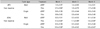
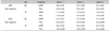
Forty three subjects satisfied the conditions and initially participated in the experiment, but 5 of them stopped using chlorhexidine and 8 were not followed up to the end of recall checks. Therefore, the final number of subjects was 30. The treatment group was determined according to the patients' preference. The cSRP group (15 subjects, mean age: 46) was used as the control group and the Fdis group (15 subjects, mean age: 51) was used as the experimental group. Table 1 shows the demographic details of both groups. This study protocol was approved by the Ethical Committee of clinical experiments, Chonnam National University Hospital (IRB No. 1-2007-06-053) and all patients provided informed consent.
2. Treatment
One week after the initial clinical examination, all the patients received supragingival scaling. One week later, the patients were divided into the control and experimental group according to their preferences and received treatment as follows. The experimental group received a modified protocol of Quirynen et al.6); 30 seconds of mouth-rinsing with a 0.1% chlorhexidine solution (Hexamedin, Bukwang, Seoul, Korea) before the procedure and an additional 10 seconds of pharynx-rinsing by bending the neck backward to hold the solution in the pharyngeal area. The patients were instructed to brush the dorsal surface of their tongues for 60 seconds. Full-mouth SRP was carried out twice in 24 hours under local anesthesia using curettes and ultrasonic instrument. After the each completion of each SRP, subgingival irrigations with 0.1% chlorhexidine solution were performed within three times for 10 minutes. Over a two week period after the procedure, the patients were instructed to perform a pharynx-rinse twice daily for 30 seconds. In order to improve the cooperation, the patients were encouraged to record the daily mouth-washing time. The control group, after one week of supragingival scaling, received subgingival SRP b y quadrants with one week intervals under local anesthesia using curettes and ultrasonic instrument. For both groups, tooth brushing instruction and oral prophylaxis were carried out at the baseline, three months and six months after the upper right quadrant SRP.
The clinical indices were measured one week after SRP, before subgingival SRP (baseline), and at o ne month, three months and six months after Fdis or cSRP at the upper right quadrant. Considering the accessibility of repeated measurement, the sulcus bleeding index (SBI), gingival recession (GR), probing depth (PD) and clinical attachment level (CAL) were measured at 6 sites in each tooth of the upper right quadrant. Overall, 42 sites per patient were measured. The plaque index (PI) was measured at 4 sites in each tooth of the upper right quadrant including the bucco-lingual and mesio-distal surfaces, making 28 sites per patient.
The sulcus bleeding index17) was measured by observing the bleeding within 30 seconds and was recorded as follows: 0 for no bleedingat all, 1 for some bleeding without gingival discoloration or swelling, 2 for bleeding and discoloration without swelling, 3 for bleeding, gingival discoloration and swelling, 4 for bleeding, gingival discoloration, swelling and ulceration, 5 for spontaneous bleeding, discoloration, distinct swelling and ulceration. The Silness & Löe index18) was used for the plaque index as follows: 0 for no plaque, 1 for a slight plaque film around the gingival margin that could be approved by explorer scratching, 2 for an excessive amount of plaque that could be observed along the marginal ridge but not in the interproximal area, 3 for an excessive amount of plaque in the marginal ridge and interproximal area. The gingival recession was measured from the CEJ to the free gingival margin using a probe (Williams probe, Hu-Friedy, Chicago, USA) using a 1 mm unit and the probing depth was also measured from the free gingival margin to the pocket base using a 1 mm unit with the same instrument. The clinical attachment level was the distance between the CEJ and pocket base, which is represented as the sum of the gingival recession and probing depth.
Adverse effects, such as an increase in body temperature, and herpes labialis were determined from a questionnaire given one week after the procedure in the upper right quadrant.
3. Statistical Methods
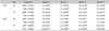
One examiner took charge of the clinical oral examination for all patients. The intra-examiner repeatibility for three patients was 99.2% for the sulcus bleeding index, 98.8% for the plaque index, 98.4% for gingival recession, 99.0% for the probing depth. All the clinical indices were recorded as the average and standard deviation for each patient by period according to the treatment method. SPSS 15.0 (SPSS Inc. USA) was used for statistical analysis. Repeated measures ANOVA was used to determine the changes in the clinical studies in 1, 3 and 6 months for each group. A t-test was used for the group difference in the changes at a specific point (1, 3, 6-month) if the measured values showed a normal distribution. A Mann-Whitney inspection was used if the values showed an abnormal distribution. A P value < 0.05 was considered significant.
RESULTS
1. Changes of clinical indices by the periods
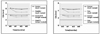
There were significant decreases in the sulcus bleeding index, plaque index and probing depth, and a significant increase in gingival recession as well as significant attachment gain at six months after both treatments (Table 2).
There was a considerable decrease in the sulcus bleeding index one month after cSRP and a slightly larger decrease until 6 months in the control group. In the experimental group the sulcus bleeding index decreased to 3 months but increased slightly by 6 months. The sulcus bleeding index in the control and experimental groups decreased to 0.9 and 1.7 at 1 month , 1.0 and 1.8 at 3 months, and 1.0 and 1.7 at 6 months, respectively (Table 3).
In the control group, there was a decrease in the plaque index until 3 months but an increase at 6 months. In the experimental group, there was a significant decrease at 1 month, as light increase at 3 months and significant increase at 6 months. The plaque index in the control and experimental groups, when compared with the values at the baseline, decreased by 0.9 and 1.9 at 1 month, 1.1 and 1.8 at 3 months, and 0.8 and 1.1 at 6 months, respectively (Table 3).
Gingival recession showed a significant increase in the control group at 1 month leading to a constant increase at 6 month. In the experimental group, there was an increase in gingival recession at 1 month, which remained constant for the remainder of the period. In the control group, gingival recession, when compared with the baseline values, increased 0.7±0.14 mm, 0.8±0.15 mm, 0.9±0.15 mm at 1, 3 and 6 months, respectively. In the experimental group, it increased 0.4±0.19 mm at one-month and remained constant until 6 months (Table 3).
In the control group, the probing depth decreased until 3 months but increased slightly at 6 months. In the experimental group, the probing depth decreased considerably at 1 month and was sustained until 6 months. In the control group, when compared with the baseline value, there was by 0.9±0.36 mm, 1.1±0.43 mm, 1.0±0.42 mm decrease observed at 1, 3 and 6 months after the procedure, respectively. In the experimental group, there was decrease of 1.2 mm until 6 months and remained constant thereafter (Table 3).
There was attachment gain of 0.1±0.36 mm in the control group, and 0.7±0.36 mm in the experimental group at 6 months, and the attachment gain was sustained for 3 months in both groups (Table 3).
There were significant decreases in the SBI and PI, significant increases in gingival recession (P<0.05), and significant attachment gain at 1 and 3 months (P<0.05). However, at 6 months, only the experimental group showed significant attachment gain (P<0.05, Table 2). When the clinical indices were compared according to period, the experimental group showed a significantly larger decrease in the SBI and PI at 1, 3 and 6 months, less gingival recession and significant attachment gain than the control group (P<0.05). On the other hand, there was similar pocket reduction in the control and the experimental group during the experiment period (Table 3).
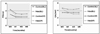
2. Changes in the probing depth and level of clinical attachment according to the initial probing depth
In the control group, the average probing depth in the area of an initial probing depth of 4~6 mm decreased from 4.7±0.24 mm to 3.4±0.17 mm and remained constant to 3.3 mm after 3 months. In the area of an initial probing depth >7 mm in the control group, the average probing depth decreased to 5.1±0.91 mm at 1 month and to 4.5±0.72 mm at 3 months but there was a slight increase to 4.8±0.94 mm at 6 months (Table 4). In the experimental group, the average pocket depth in the area of initial probing of 4~6 mm decreased from 4.6±0.13 mm at the baseline to 3.0±0.22 mm at 1 month and remained constant to 2.9 mm after 3 months. In the area of an initial probing depth >7 mm in the experimental group, the probing depth decreased to 4.5±0.61 mm at 1 month and 4.3±0.47 mm at 3 months but slightly increased to 4.4±0.52 mm at 6 months (Table 4).
In the control group, the level of clinical attachment in the area of an initial depth of 4~6 mm increased from 5.4±0.42 mm to 4.9±0.57 mm at 1 month and 4.8±0.57 mm at 3 months but decreased slightly to 5.0±0.63 mm at 6 months. In the area of an initial pocket depth >7 mm in the control group, the level of attachment increased from 8.7±1.49 mm to 7.3±2.20 mm at 1 month, 6.9±2.03 mm at 3 months, but decreased slightly to 7.2±2.23 mm at 6 months. In the experimental group, the level of attachment in the area of an initial pocket depth of 4~6 mm showed a steady increase from 5.0±0.41 mm to 3.9±0.45 mm until 6 months. In addition, in the area of an initial pocket depth >7 mm in the experimental group, there was an increased from 8.3±1.75 mm to 6.0±1.46 mm at 1 month, 6.0±1.24 mm at 3 months but a slight decrease to 6.1±1.31 mm at 6 months (Table 4).
In both groups, the pocket reduction and clinical attachment gain in the area of the initial pocket depth of 4-6 mm and >7 mm were significant until 6 months compared with the baseline (P<0.05, Table 4).
When comparing the clinical indices according to the period, the pocket reduction and clinical attachment gain in the area of an initial probing depth of 4-6 mm in the experimental group was significantly larger than the control group until 6 months (P<0.05). However, there were no significant differences between the groups in the area of a probing depth >7 mm (Table 5).
5. Adverse effects after treatment
A questionnaire was given to the patients after one week of treatment in the upper right quadrant to determine if there were any adverse effects after treatment. In the experimental group, one patient reported an increase in body temperature and three patients with a prior medical history reported herpes labialis. However, there were no adverse effects reported in the control group.
DISCUSSION
Pathogenic microbes can re-establish in the dorsum of the tongue, mucosa, tonsil and saliva as well as in the gingival pocket after periodontal treatment. Moreover, those microbes can be transmitted via saliva flow19), periodontal probe20), explorer21) and even by oral hygienic instruments22). Therefore, in the case of non-surgical periodontal treatment, access to the oral cavity as a whole might be more reasonable than access in the part. In addition, antiseptics may also be used to effectively suppress cross contamination. Since the introduction of Fdis by Quirynen et al6), this idea of access as a whole has proven to be more effective clinically and microbiologically than the conventional way of partial access in many studies. However, chlorhexidine gel, spray and solution (0.2%) are not available in Korea. Therefore, in this study of generalized moderate to severe chronic periodontitis patients, a chlorhexidine solution (0.1%), which can be purchased over-the-counter and has few side-effects, was used in the modified Fdis that was designed to be simple to apply after SRP. In addition, the clinical effects of Fdis were compared with the conventional SRP over a 6-month period.
Although the additional effects of chlorhexidine use cannot be excluded, a full-mouth root planing (Frp) group, which carried out SRP without using chlorhexidine within 24 hours, was excluded from this experiment in order to compare the effect. A previous study reported no significant clinical and microbiological difference between Frp and Fdis but early stage healing i.e. accelerated pocket reduction and clinical attachment, was more advantageous after Fdis7). In addition, a preliminary study reported that Fdis is particularly beneficial in the short-term follow-up with an immediate effect on pocket reduction and clinical attachment gain23). The major effect of Fdis is associated with the completion of instrumentation within 24 hours, which means the untreated pockets are a major pathogenic microbial reservoir with other ecological niches making a minor contributor24). In addition, the effect of Fdis can be interpreted as a local Schwartzman reaction, which is the induction of an antibody from the release of a large number of antigens after instrumentation25). However, Apatzidou et al11) reported no significance in this vaccination.
This study reported that SBI and PI were reduced leading to more effective removal of gingival inflammation and plaque in the early stages after the modified Fdis than the conventional method. Even when the measured PI was higher after Fdis than the conventional method, the gingival index was lower after Fdis than the conventional method, which means that Fdis is more effective in controlling gingival inflammation9). The level of plaque deposition decreased significantly in the modified Fdis group when using the chlorhexidine solution, which is effective in controlling plaque in the early stages of healing. However, after considering the significant increase in plaque deposition after quitting chlorhexidine use in the experimental group compared with the control group, either the period of chlorhexidine use needs to be extended or oral hygiene needs to be emphasized to the patients through regular check-ups.
In this study, the probing depth in the area of the initial pocket depth of 4~6 mm decreased 1.4 mm after cSRP and 1.7 mm after Fdis. Knöfler et al.14) reported a probing depth in the area of the initial pocket depth of 4~6 mm, which decreased by 1.1 and 1.0 mm after cSRP and Fdis, respectively. Jervoe-Storm et al26) reported similar results showing decrease of 1.6 mm and 1.5 mm, respectively. In this study, the deep (≥7 mm) initial pockets decreased by 3.0 and 3.3 mm after cSRP and Fdis, respectively, which shows a far larger decrease than the area of the moderately deep (4-6 mm) initial pockets. Fdis showed far more pocket reduction than cSRP. However, Fdis showed a significant difference in the decrease in the probing depth and attachment gain in the moderately deep (4-6 mm) initial pockets than in the deep (≥7 mm) initial pockets compared with the result from cSRP. These results are not in accordance with other studies, which reported a larger significant clinical difference in the deep (≥7 mm) initial pockets than in the moderately deep (4-6 mm) initial pockets6,27). The different effects of Fdis in the previous study were due not only to the form of chlorhexidine and its concentration, but also to the accessibility of the antiseptics to the deepest part of the pocket because a syringe with a blunt needle was used to the point of feeling resistance in the subgingival irrigation. Therefore, more study on the modified Fdis will be needed to determine the additional clinical effects with using these instruments in the deep pocket.
When comparing the change in probing depth and attachment level according to tooth type, multi-rooted teeth showed much more clinical improvement after the two methods than single-rooted teeth as the initial probing depth was deeper in the multi-rooted teeth. By comparing the effect of the two methods according to the period, single-rooted teeth showed little difference in pocket reduction and attachment gain. Therefore, single-rooted teeth can be also treated effectively by cSRP. Multi-rooted teeth showed significantly larger attachment gain after modified Fdis. This is thought to be due to the additional use of antiseptics to the area with limited accessibility in the sight and instrumentation resulting in improved healing in the early period.
A comparison of the difference in pocket reduction and attachment gain by the bucco-lingual and proximal surface revealed the proximal surface to show more improvement after the two methods than the bucco-lingual surface because the initial depth on the proximal surface was deeper. A comparison of the treatment effects of the two methods according to the period, revealed no significance in pocket reduction between the two methods. However, there was a significant difference in attachment gain at 1 and 6 months in the bucco-lingual surface and at 1, 3 and 6 months in the proximal surface after Fdis. There was significantly less gingival recession after Fdis compared with cSRP. Bollen et al8) reported 1.4 mm less gingival recession after Fdis than after cSRP. Therefore, the healing process of Fdis is more advantageous to gingival recession and clinical attachment gain.
The reported effects of Fdis are not consistent, and some parts of this study showed little improvement compared with the conventional SRP. However, overall, Fdis was reported to show more improvement than cSRP. The reason for this difference between Fdis and cSRP can be found in the procedural protocol, particularly in the aspect of the risks of cross contamination28). The risk of cross contamination is related to many factors, such as the protocol for applying chlorhexidine, area of application, concentration, period of usage, treatment interval, oral hygiene, the severity of periodontitis, the amounts of plaque before treatment, and the care after treatment. In previous studies, SRP was administered at two week intervals, which increased the risk of cross-contamination. Knöfler et al. administered SRP on 2 quadrants at 4 weeks' interval, which increased the risk of cross contamination14). However, there were no significant differences in the clinical effects between SRP and Fdis. In this study, supragingival full-mouth scaling was administered in advance. The subgingival treatment was performed one week after scaling while cSRP was carried out by quadrant at one-week intervals. Fdis showed significant improvement compared with cSRP. This difference may be due to the time of the probing depth measurement. While the baseline probing depth was measured before treatment in this study, in some studies reporting significant decrease in probing depth, the baseline probing depth was measured after supragingival scaling in order to reduce the measurement error from the calculus. As a result, the probing depth may be measured deeper at the baseline, showing an exaggerated decrease in the probing depth from Fdis.
Fdis has the advantages of reducing the treatment time and cost in addition to its beneficial clinical and microbiological effects. In this study, all patients were divided into a control and experimental group according to their preferences. Most of them preferred Fdis. In addition, Fdis has its advantage in medication. Administering cSRP to a patient who is taking daily anti-coagulants can double the non-surgical periodontal treatment period compared with a healthy patient. However, Fdis reduced the time for abstaining from the anti-coagulant, which decreased the systemic risk caused by the discontinuation of medication. Fdis also has an advantage to systematically healthy periodontitis patients. In this study of Fdis, medication was prescribed only once by instructing the patients to take antibiotics and antiphlogistic agent for only 3 days while medication was prescribed on the completion of each quadrant in cSRP, which increases the time and cost of medication.
Some authors reported adverse effects of Fdis, such as increased body temperature, herpes labialis, etc. However, in this study, only one patient reported increased body temperature and three patients reported herpes labialis with a prior medical history. These side effects may result from the supragingival scaling, which decreased the number of microbes in the subgingival area before subgingival SRP and time of exposure to mechanical stress. Another cause is believed to be the antibiotics and anti-inflammatory agents used for 3 days. There was a study reporting the relief of the increased body temperature after Fdis when azithromycin was administered systemically before the treatment of severe periodontitis patients29). These side effects can be minimized if efforts are made to reduce the iatrogenic trauma. Overall, this study revealed modified Fdis is more effective than cSRP 6 months after treatment for generalized moderate to severe chronic periodontitis.
Fdis is particularly effective when the risk of cross contamination is high as a result of inadequate plaque control and the massive deposition of plaque and calculus in the untreated areas. It is also advantageous when patients cannot visit clinics repeatedly for financial reasons and schedule or when the patients are taking anti-coagulants, which can prolonged the treatment time if the conventional method is used or have systemic diseases that retard the healing procedure. The proper administration of modified Fdis would ensure improved treatment results particularly when the earlier healing is needed for patients scheduled to undergo radiation therapy.
This study compared the clinical effects of modified Fdis after scaling using a chlorhexidine solution (0.1%) with those of conventional SRP in the treatment of moderate to severe generalized chronic periodontitis after a 6 month follow-up. After Fdis for the treatment of moderate to severe generalized chronic periodontitis, there was a reduction of gingival inflammation, plaque level, and probing depth, a smaller increase in gingival recession, and attachment gain in the teeth with an initial pocket depth of a moderate level (4~6 mm) and multi-roots, and larger attachment gain in the proximal surface compared with the bucco-lingual surface. Within the limitations of this study, modified Fdis has more beneficial effects in the treatment of moderate to severe generalized chronic periodontitis after 6 months than conventional SRP.


 XML Download