

 PDF
PDF ePub
ePub Citation
 Print
Print


INTRODUCTION
The main objective of periodontal treatment is not only to relieve symptoms but also to regenerate the destroyed tissues. Many methods have been introduced for regenerating damaged periodontal tissues. Guided tissue regeneration (GTR) therapy was introduced in the 1980s to achieve a repopulation of the periodontal ligament fibroblasts, and was shown to promote periodontal regeneration. The membrane barrier used in GTR should be histocompatible, biocompatible and have the capacity for space maintenance1).
Absorbable membrane does not require a second surgical procedure, and membrane exposure is rare. Aquirre et al2) showed new bone formation in bony defects using absorbable membrane. However, controlling the time of absorption is difficult and therefore could cause a localized inflammatory reaction. In addition to the above disadvantages, poor membrane stability in the wet state causes space loss between the tooth and the membrane, producing poor clinical results3). An absorbable membrane should be used in GTR in places where exact initial closure is possible, since complete removal is difficult when exposed4). To solve this problem, many studies have been carried out on biodegradable membranes, and acceptable results were presented5). Theideal membrane should be absorbable and should not require removal after the tissue regenerates; it also should block tissue migration effectively and resist inflammatory reaction. Lastly, it must have space-maintaining capacity.
Recently, interest in chitosan has increased due to its excellent biological properties such as biocompatibility, antibacterial effect, and rapid healing capacity. Chitin and chitosan (poly-N-acetyl glucosaminoglycan), a carbohydrate biopolymer extracted from chitin, are the second most abundant natural biopolymers next to cellulose. Chitin is a primary structural component of the exoskeleton of arthropods (e.g. crustaceans), the cell wall of fungi, and the cuticle of insects. Chitin is a very stable polysaccharide and is a linear polymer of N-acetyl-D-glucosamine monomers joined in a 1,4β-glucosidic linkage. Chitosan is a derivative of chitin obtained by N-acetylating chitin. As with polymers in general, enzymes can hydrolyze chitin and chitosan. The most effective enzyme for this process is lysozyme6,7).
Although the healing effects of chitin and chitosan on mammalian wounds have been known for centuries, it was not until the 1960s that the ingredients were documented8).
Other studies since have suggested that chitosan enhances the formation of bone tissue and it could be used as the matrix of tissue engineering for gingiva9). Paik et al10) reported that chitosan enhanced type I collagen synthesis in the early stage, and facilitated differentiation into osteogenic cells in the human periodontal ligament fibroblasts in vitro. In addition, a chitosan/collagen sponge applied to one-wall intrabony defects surgically created in beagle dogs inhibited the apical migration of the epithelium and enhanced the growth of new bone and new cementum11).
Another biomaterial of interest is hydroxyapatite, which is a major component of human bone. Hydroxyapatite is used as bone substitute in the fields of orthopedics and dentistry because of its good osteoconductivity, bioactivity and biocompatibility12). But hydroxyapatite is brittle and easy to fracture so it is difficult to mould into a specific shape. The hydroxyapatite-chitosan (HA-CS) complex, containing hydroxyapatite nanoparticles, was therefore developed to overcome the original disadvantage of hydroxyapatite13).
Although many materials are used to regenerate periodontal tissues, there is as yet no material that satisfies all conditions. This paper reports on the fabrication of the HA-CS membrane in diverse proportion. The objective of this study was to evaluate bone regeneration capacity of HA-CS membrane in rat calvarial defects.
MATERIALS AND METHODS
This study included 70 male Sprague-Dawley rats (body weight 250-300 g) maintained in plastic cages in a room with a 12 hour day/night cycle and an ambient temperature of 21 ℃. The rats were allowed free access to water and standard laboratory fool pellets. Animal selection, management, surgical protocol, andpreparation followed the routines approved by the Institutional Animal Care and Use Committee, Yonsei Medical Center, Seoul, Korea(approval No.: 06-171).
Chitosan was dissolved in a 2% acetic acid solution, and then mixed with phosphoric acid solution. After Ca(OH)2 was added to this solution, HA was able to be synthesized. At that time, HA proportion was regulated, and 0:100, 30:70, 40:60, 50:50 (HA weight: CS weight) HA-CS composites were developed. After citric acid was added to each HA-CS composite, original solution for threading was developed. The HA-CS solution was filtered and threaded in a 10% NaOH solution and then washed and dried to develop the HA-CS membrane. Some of the membrane, HA 30%/CS 70% and HA 50%/CS 50%, were pressed for advanced mechanical properties. The size of HA nanoparticles was 20-100 nm. All HA-CS membranes passed the cytotoxic test, MTT assay.
Eight-millimeter critical-sized calvarial defects were created in 70 male Sprague-Dawley rats. The animals were divided into 7 groups of 10 animals and received either 1) chitosan (CS) 100% membrane, 2) hydroxyapatite (HA) 30%/CS 70% membrane, 3) HA 30%/CS 70%, pressed membrane, 4) HA 40%/CS 60% membrane, 5) HA 50%/CS 50% membrane, 6) HA 50%/CS 50%, pressed membrane, or 7) a sham - surgery control.
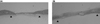
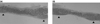
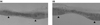
The animals were sacrificed by CO2 asphyxiation at 2 or 8 weeks post-surgery. The samples were fixed and decalcified and embedded in paraffin, stained with hematoxylin/eosin (H-E), and examined with an optical microscope. The sections were examined at 20x magnification for histometric evaluation. Measurements included % defect closure, % augmented bone area, and % new bone area (Fig. 1). Histometric recordings from the samples were used to calculate mean and standard deviations. We used one-way ANOVA, Least Significant Difference (LSD) method and paired t-test for statistical analysis.
RESULTS
1. Clinical observations
Wound healing was generally uneventful and appeared similar for all nonpressed-membrane experimental groups (Exp. 1, 2, 4 and 5). In some pressed-membrane experimental groups (Exp. 3 and 6), slight inflammatory reaction was observed at 2 weeks, but inflammation was not detected at 8 weeks. Material exposure or other complications of the surgical sites were not observed.
2. Histologic observations
1) Control
At 2 and 8 weeks post-surgery, defects filled with thin, loose connective tissue, with minimal new bone formation originating from the defect margins, were observed. The defect center had collapsed (Fig. 2).
2) Experimental groups
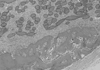
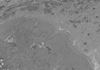
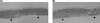
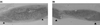
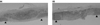
In both the chitosan-only and HA-CS membrane groups, the defects were filled with loose or dense, fibrous connective tissue, and limited new bone formation was observed at the defect margins at 2 weeks. A large number of residual chitosan fibers and hydroxyapatite particles were observed within the new bone at 2 weeks (Fig. 3), but there appeared to be fewer of these at 8 weeks (Fig. 4). Irrespective of the hydroxyapatite and chitosan dose levels, all defect sites exhibited bone formation, and volume was increased. At 8 weeks, the appearance of the new bone was more lamellar than that observed at 2 weeks.
Membrane remnants are composed of chitosan fibers and hydroxyapatite particles. The membrane remnants were surrounded by connective tissue. As the HA dose of membrane increased, the size of membrane remnants had decreased at 8 weeks.
3. Histometric analysis
Three animals were excluded from the histometric analysis due to technical complications in the histologic processing. Only limited new bone formation was observed in the controls. Irrespective of the HA-CS dose, there were no significant differences in % defect closure at either 2 or 8 weeks post-surgery. There was also no statistically significant difference between the results at 2 and 8 weeks in terms of % defect closure. However, one-way ANOVA revealed that new bone and augmented areas did show significant differences in each healing interval (p<0.01).
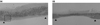
Tables 1 and 2 show the results of histometric analysis. New bone deposition between 2 and 8 weeks was significantly different in the Exp. 1 and Exp. 2 groups (Fig. 5, 6). Augmented areas were significantly decreased from 2 weeks to 8 weeks post-surgery in the pressed-membrane group (Fig. 7, 8, 9, 10).
New bone formation for the 50% HA dose groups (Exp. 5 and Exp. 6) at 2 weeks post-surgery was significantly greater than for the HA 0% group (Exp. 1) or HA 30% group (Exp. 2). There were significant differences between 2 and 8 weeks in new bone formation in HA 0% group (Exp. 1) and HA 30% group (Exp. 2). But Exp. 5 and Exp. 6, which received the high HA dose, were not statistically significant.
DISCUSSION
Many absorbable membranes made of collagen or various kinds of polymers, including chitosan, have been developed to the present day, and many studies on their healing effects have been carried out14-17).
Chitosan is known to accelerate cell migration and tissue maturation, leading to wound healing; therefore, many studies relating to this property are being undertaken in the fields of dentistry and orthopedic surgery. Chitosan could be adhesive to bioactive materials such as PDGF and BMP, and thus could be widely used clinically in addition to bone substitutes and barriers18). The nano type of hydroxyapatite can also be attached to chitosan fibers.
Nano-sized HP may have other special properties due to its small size and huge specific surface area. Webster et al19) have demonstrated a significant increase in protein adsorption and osteoblast adhesion on the nano-sized ceramic. Studies have shown that nano-HA/chitosan composite scaffolds may serve as a good three-dimensional substrate for cell attachment in vitro and migration in engineered bone and periodontal tissue20). Some researchers experimented with a composite of chitosan and nano-HA paste, but it did not have porosity and could not be loaded with cells. Others have reported that fibrous scaffolds had a much greater surface-to-volume ratio than scaffolds with solid pore walls, which might have further increased protein adsorption capacity21,22).
In this study, we attempted to show the clinical efficacy of a newly-formed fibrous hydroxyapatitechitosan (HA-CS) membrane in a rat model. The experimental model used in this study has been shown to be effective for evaluating the potential for bone formation23-25). The rat calvarial defect model is convenient for examining bone regeneration because of its effective accessibility and lack of fixation requirements.
In our histometric analysis, the length and the area of the new bone formation were compared. Measurements were taken by using computer software, called Image Pro Plus. Specimens were obtained from the middle coronal section. The length of new bone formation was measured to compare the amount of cell migration. The more the cells migrate, the higher the possibility of bone union. As the cells' length growth increases, in considering the thickness, more bone formation could be predicted. Therefore, this could be regarded to be a good marker for the membrane's bone regenerative capacity. Many rat studies have shown a significant increase in the area and density of newly formed bone when water-soluble oligo-chitosan was applied to a calvarial defect26) and chitosan reconstituted with absorbable collagen sponge has significant potential to induce the regeneration of bone in rat calvarial critical-sized defects27). Previous study showed augmented area, including new bone, of chitosan/absorbable collagen sponge group in 2 weeks post-surgery was 6.19 ± 2.03 mm2 and 8 weeks post-surgery was 4.84 ± 0.88 mm2. All of experimental groups were enhanced augmented area both 2 and 8 weeks post-surgery in present study.
As the HA dose increased, there was more new bone formation in the early healing period, 2 weeks after surgery, while there was no significant difference as time went on. This suggests that HA nanoparticles may resorb quickly and induce new bone in the early stage of healing.
Ideally, the bone substitute should conduct or induce bone formation at the same time as it is completely resorbed and substituted by bone tissue. Evidence from previous studies suggests that HA resorption can be mediated by cells or by disintegration through the action of extracellular fluids28,29). Our histologic study also showed the presence of multinuclear giant cells in close contact with the HA and chitosan surface and bone formation adjacent to the particles. Liljensten et al30) stated that even for HAs considered absorbable, the resorption process is slow and its finalization is not well determined.
Levels of wound healing and bone formation were similar in the non-pressed membrane experimental groups. But in some pressed-membrane experimental groups, inflammation was observed at 2 weeks after surgery but had subsided at 8 weeks. Andrade et al31) studied denseand porous HA cylinders and observed the fibrous tissue development surrounding dense implants and the direct contact of the new bone formed in the porous implants. Takeshita et al32) used dense HA granules (300 to 600µm) in bone defects created around osseointegrable implants and reported fibrous encapsulation of the granules. They concluded that dense HA granules negatively interfered with bone formation. Many other studies have reported improved bone-HA integration when the particles presented micro- and macropores. Our study also suggests that pressed-membrane with high proportion of HA particles in a unit area could cause inflammation in early healing period.
The augmented area of the pressed-membrane groups (Exp. 3 and Exp. 6) was significantly decreased after 8 weeks. As the HA dose increased, the augmented areas decreased at 8 weeks because the HA nanoparticles were absorbed earlier than the chitosan fiber.
From a histological view, it seems that the membrane lacked any major role as a scaffold. There was no statistical significance in defect closure that we could evaluate from length-growth of cells. Therefore, it appears that the HA-CS membrane does not stimulate cell migration to the center of defect. In summary, HA-CS membrane could collapse early in the healing period and seems to interfere in the formation of new bone in the central zone of surgical defects.
The pressed-membrane groups were developed primarily to overcome the weak mechanical properties of the conventional membranes and to be easy to handle. After the pressed-membrane absorbed water, however, there was no difference in handling from non-pressed, conventional membranes. Furthermore, pressed-membrane showed swollen shape at 2 weeks after surgery. To play a leading role in a scaffold, membranes should have improved mechanical properties in terms of water absorption.
In summary, surgical implantation of the HA-CS membrane resulted in enhanced local bone formation at both 2 and 8 weeks compared to the control group. The results suggest that the addition of HA accelerated new bone formation as seen in Table 3. Further studies will be required to improve the mechanical properties for development of a more rigid, pressed scaffold for the HA-CS membrane.

 XML Download