

 PDF
PDF ePub
ePub Citation
 Print
Print


Kuk-Bong Cha1, Won-Kyung Kim2, Young-Kyoo Lee2, Young-Sung Kim2
Abstract
Purpose
Ultrasonic scalers have been widely used for removing biofilm which is considered as major etiologic factor of periodontal disease. The purpose of this study was to evaluate the effect of working parameters of piezoeletric ultrasonic scaler with scaler tip (No. 1 tip) on casting gold alloy removal.
Methods
Type III dental casting gold alloy (Firmilay ® , Jelenko Inc, CA, USA) was used as substitute for tooth substance. Piezoeletric ultrasonic scaler and No.1 scaler tip (P-Max ® , Satelec, France) were selected. The selected working parameters were mode (P mode, S mode), power setting (2, 4, 8) and lateral force (0.5 N, 1.0 N, 2.0 N). The effect of working parameters was evaluated in terms of ablation depth, ablation width and ablation area.
Results
Mode influenced ablation depth and ablation area. Power also influenced ablation depth and ablation area. Especially, Power 2 and power 8 showed statistically significant difference. Lateral force had influence on ablation width, and 0.5 N resulted significant increase compared with 1.0 N and 2.0 N. Ablation depth was influenced by mode, power and lateral force and defect width was influenced by lateral force. Ablation area was influenced by mode and power.
Figures and Tables
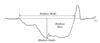
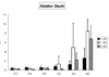
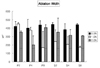
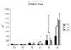
Figure 5
Influence of working parameters on ablation depth in terms of quadratic interactions showing significance. Horizontal bar indicates statistically significant combination from general linear models procedure of least squares means at significance level 0.05.
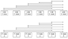
Figure 6
Influence of working parameters on ablation width in terms of quadratic interactions showing significance. Horizontal bar indicates statistically significant combination from general linear models procedure of least squares means at significance level 0.05.
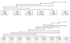
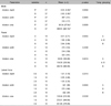
Figure 7
Influence of working parameters on ablation area in terms of quadratic interactions showing significance. Horizontal bar indicates statistically significant combination from general linear models procedure of least squares means at significance level 0.05.

References
1. Löe H, Theilade E, Jensen SB. Experimental gingivitis in man. J Periodontol. 1965. 36:177–187.

2. Costerton JW, Stewart PS, Greenberg EP. Bacterial biofilms: acommon cause of persistent infections. Science. 1999. 284:1318–1322.
3. Darveau RP, Tanner A, Page RC. The microbial challenge in periodontitis. Periodontol 200. 1997. 14:12–32.
4. Socransky SS, Haffajee AD. Dental biofilms: difficult therapeutic targets. . Periodontol 200. 2002. 28:12–55.
5. Stende GW, Schaffer EM. A comparison of ultrasonic and hand scaling. J Periodontol. 1961. 32:312.
6. Gellin RG, Miller MC, Javed T, Engler WO, Mishkin DJ. The effectiveness of the Titan-S sonic scaler versus curettes in the removal of subgingival calculus: a human surgical evaluation. J Periodontol. 1986. 57:672–680.
7. Breininger DR, O'Leary TJ, Blumenshine RV. Comparative effectiveness of ultrasonic and hand scaling for the removal of subgingival plaque and calculus. J Periodontol. 1987. 58:9–18.
8. Caffesse RG, Alspach SR, Morrison EC, Burgett FG. Scaling and root planning with and without periodontal flap surgery. J Clin Periodontol. 1986. 13:205–210.
9. Buchanan SA, Robertson PA. Calculus removal by scaling/root planningwith and without surgical access. J Periodontol. 1987. 58:159–163.
10. Matia JI, Bissada NF, Maybury JE, Riccetti P. Efficiency of scalng of the molar furcation area with and without surgical access. Int J Periodontics Restorative Dent. 1986. 6:25–35.
11. Fleischer HC, Mellonig JT, Brayer WK, Gray JL, Barnett JD. Scaling and root planning efficacy in multirooted teeth. J Periodontol. 1989. 60:402–409.
12. Parashis AO, Anagnou-Vareltzides A, Demetriou N. Calculus removal from multirooted teeth with and without surgical access. I. Efficacy on external and furcation surfaces in relation to probing depth. J Clin Periodontol. 1993. 20:63–68.
13. Zappa U, Smith B, Simona C, Graf H, Case D, Kim W. Root substance removal by scaling and root planning. J Periodontol. 1991. 62:750–754.
14. Fogel HM, Pashley DH. Effect of periodontal root planning on dentin permeability. J Clin Periodontol. 1993. 20:673–677.
15. Fukazawa E, Nishimura K. Superficial cemental curettage: its efficacy in promoting improved cellular attachment on human root surfaces previously damaged by periodontitis. J Periodontol. 1994. 65:168–176.
16. Kerry GJ. Roughness of root surfaces after use of ultrasonic instruments and hand curettes. J Periodontol. 1967. 38:340–346.
17. Wilkinson RF, Maybury JE. Scanning electron microscopy of the root surface following instrumentation. J Periodontol. 1973. 44:559–563.
18. Aleo JJ, De Renzis FA, Farber PA. In vitro attachment of human gingival fibroblasts to root surface. J Periodontol. 1975. 46:639–645.
19. Nakib NM, Bissada NF, Simmelink JW, Goldstine SN. Endotoxin penetration into root cementum of periodontally healthy and diseased human teeth. J Periodontol. 1982. 53:368–378.
20. Checchi L, Pelliccioni GA. Hand versus ultrasonic instrumentation in the removal of endotoxins from root surfaces in vitro. J Periodontol. 1988. 59:398–402.
21. Cheetham WA, Wilson M, Kieser JB. Root surface debridement - an in vitro assessment. J Clin Periodontol. 1988. 15:288–292.
22. Smart GJ, Wilson M, Davies EH. The assessment of ultrasonic root surface debridement by determination of residual endotoxin levels. J Clin Periodontol. 1990. 17:174–178.
23. Claffey N, Polyzois I, Ziaka P. An overview of nonsurgical and surgical therapy. Periodontol 200. 2004. 36:35–44.
24. Trenter SC, Walmsley AD. Ultrasonic dental scaler: associated hazards. J Clin Periodontol. 2003. 30:95–101.
25. Arabaci T, Çiçek Y, Çanakçi CF. Sonic and ultrasonic scalers in periodontal treatment: a review. Int J Dent Hyg. 2007. 5:2–12.
26. Drisko CH. Root instrumentation: power-driven versus manual scalers, which one? Dent Clin North Am. 1998. 42:229–244.
27. Flemmig TF, Petersilka GJ, Mehl A, Hickel R, Klaiber B. The effect of working parameters on root substance removal using a piezoelectric ultrasonic scaler in vitro. J Clin Periodontol. 1998. 25:158–163.
28. Lee Young-kyoo. Root surface roughness following mechanical instrumentation in vivo and in vitro SEM study. J Korean Acad Periodontol. 1998. 28:823–828.
29. Lee Young-kyoo. The Effect of a Piezoelectric Ultrasonic Scaler with Curette Tip on Root Substitute Removal in Vitro. J Korean Acad Periodontol. 2000. 30:429–441.
30. Lee Young-kyoo. The Effect of a Piezoelectric Ultrasonic Scaler with Curette Tip on Casting Gold Removal in Vitro. J Korean Acad Periodontol. 2001. 31:531–542.
32. Craig RG. Restorative dental materials. 1993. 9th ed. St. Louis: Mosby-Year Book, Inc.;89.
33. Hildebrand CN, Morse DR. Periodontic-endodontic interrelationships. Dent Clin North Am. 1980. 24:797–812.
34. Nyman S, Westfelt E, Sarhed G, Karring T. The role of diseased root cementum in healing following treatment of periodontal disease. J Clin Periodontol. 1988. 15:464–468.
 XML Download