

 PDF
PDF ePub
ePub Citation
 Print
Print


Abstract
Purpose
The integrity of interproximal hard/soft tissue has been widely accepted as the key determinant for success or degree of root coverage following the connective tissue graft. However, we reason that the gingival biotype of an individual, defined as the distance from the interproximal papilla to gingiva margin, may be the key determinant that influence the extent of root coverage regardless of traditional classification of gingival recession. Hence, the present study was performed with an aim to verify that individual gingival scalloping pattern inherent from biotype influence the level of gingival margin following the connective tissue graft for root coverage.
Methods
Test group consisted of 43 single-rooted teeth from 21 patients (5 male and 16 female patients, mean age: 36.6 years) with varying degrees of gingival recession requiring connective tissue graft; 20 teeth of Miller class I and 23 teeth of Miller class III gingival recession, respectively. The control group consisted of contralateral teeth which did not demonstrate apparent gingival recession, and thus not requiring root coverage. For a biotype determination, an imaginary line connecting two adjacent papillae of a test tooth was drawn. The distance from this line to gingival margin at mid-buccal point and this distance (P-M distance) was designated as "gingival biotype" for a given individual. The distance was measured at baseline and 3 to 6 months examinations postoperatively both in test and control groups. The differences in the distance between Miller class I and III were subject to statistical analysis by using Student's t-test while those between the test and control groups within a given patient were by using paired t-test.
Results
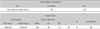
The P-M distance at 3 to 6 months postoperatively was not significantly different between Miller class I and Miller class III. It was not significantly different between the test and control group in a given patient, either, both in Miller class I and III.
Conclusions
The amount of root coverage following the connective tissue graft was not dependent on Miller's classification, but rather was dependent on P-M distance, strongly implying that the gingival biotype of a given patient may play a critical impact on the level of gingival margin following connective tissue graft.
Figures and Tables

Table 2
Comparision of P-M Distance between Miller class I and III following the Connective Tissue Graft


References
1. Santarelli GA, Ciancaqlini R, Campanari F, Dinoi C, Ferraris S. Connective tissue grafting employing the tunnel technique : a case report of complete root coverage in the anterior maxilla. Int J Periodontics Restorative Dent. 2001. 21(1):77–83.
2. Hall WB. Present status of soft tissue grafting. J Periodontol. 1977. 48(9):587–597.

3. Baker DL, Seymour GJ. The possible pathogenesis of gingival recession. A histological study of induced recession in the rat. J Clin Periodontol. 1976. 3(4):208–219.
4. Khocht A, Simon G, Person P, Denepitiya JL. Gingival recession in relation to history of hard toothbrush use. J Periodontol. 1993. 64(9):900–905.
5. Smukler H, Landsberq J. The toothbrush and gingival traumatic injury. J Periodontol. 1984. 55(12):713–719.
6. Boyd RL. Mucogingival considerations and their relationship to orthodontics. J Periodontol. 1978. 49(2):67–76.
7. Donaldson D. Gingival recession associated with temporary crowns. J Periodontol. 1973. 44(11):691–696.
8. Wilson RD. Marginal tissue recession in general dental practice: a preliminary study. Int J Periodontics Restorative Dent. 1983. 3(1):40–53.
9. Allen EP, Miller PD Jr. Coronal positioning of existing gingiva: short term results in the treatment of shallow marginal tissue recession. J Periodontol. 1989. 60(6):316–319.
10. Blanes RJ, Allen EP. The bilateral pedicle flap-tunnel technique: a new approach to cover connective tissue graft. Int J Periodontics Restorative Dent. 1999. 19(5):471–479.
11. Pennel BM, Tabor JC, King KO, et al. Free masticatory mucosa graft. J Periodontol. 1969. 40(3):162–166.
12. Miller PD Jr. A classification of marginal tissue recession. Int J Periodontics Restorative Dent. 1985. 5(2):8–13.
13. Langer B, Langer L. Subepithelial connective tissue graft technique for root coverage. J Periodontol. 1985. 56(12):715–720.
14. Roccuzzo M, Bunino M, Needleman I, Sanz M. Periodontal plastic surgery for treatment of localized gingival recessions: a systematic review. J Clin Periodontol. 2002. 29:Suppl 3. 178–194.
15. Miller PD Jr. Root coverage using the free soft tissue autograft following citric acid application. III. A successful and predictable procedure in areas of deep-wide recession. Int J Periodontics Restorative Dent. 1985. 5(2):14–37.
16. Melcher AH. On the repair potential of periodontal tissues. J Periodontol. 1976. 47(5):256–260.
17. Seibert J, Lindhe J. Esthetics and periodontal therapy. Textbook of clinical periodontology. 2nd ed. Copenhagen: Munksgaard;477–514.
18. Muller HP, Eger T. Gingival phenotypes in young male adults. J Clin Periodontol. 1997. 24(1):65–71.
19. Olsson M, Lindhe J. Periodontal characteristics in individuals with varying form of the upper central incisors. J Clin Periodontol. 1991. 18(1):78–82.
20. Claffey N, Shanley D. Relationship of gingival thickness and bleeding to loss of probing attachment in shallow sites following nonsurgical periodontal therapy. J Clin Periodontol. 1986. 13(7):654–657.
21. Sanavi F, Weisgold AS, Rose LF. Biologic width and its relation to periodontal biotypes. J Esthet Dent. 1998. 10(3):157–163.
22. Weisgold AS. Contours of the full crown restoration. Alpha Omegan. 1977. 70(3):77–89.
23. Weisgold AS, Arnoux JP, Lu J. Single-tooth anterior implant: a world of caution. Part I. J Esthet Dent. 1997. 9(5):225–233.
24. Olsson M, Lindhe J, Marinello CP. On the relationship between crown form and clinical features of the gingiva in adolescents. J Clin Periodontol. 1993. 20(8):570–577.
25. Pontoriero R, Carnevale G. Surgical crown lengthening: a 12-month clinical wound healing study. J Periodontol. 2001. 72(7):841–848.
26. Langer B, Calagna LJ. The subepithelial connective tissue graft a new approach to the enhancement of anterior cosmetics. Int J Periodontics Restorative Dent. 1982. 2(2):22–33.
27. Lindhe J, Karring T, Lang NP. Clinical Periodontology and Implant Dentistry. 2003. 4th ed. Blackwell Munksgaard;611–613.
28. Zucchelli G, Testori T, De Sanctis M. Clinical and anatomical factors limiting treatment outcomes of gingival recession: a new method to predetermine the line of root coverage. J Periodontol. 2006. 77(4):714–721.
 XML Download