

 PDF
PDF ePub
ePub Citation
 Print
Print


Abstract
Purpose
The purpose of this study is to evaluate correlation according to sites measured from women diagnosed with osteoporosis without other factors influencing osteoporosis.
Materials and Methods
Two hundred patients diagnosed with osteoporosis using dual-energy X-ray absorptiometry from January 2006 to January 2007 were evaluated. All patients were measured at the hip joint (Femur neck, Ward triangle, and great trochanter), lumbar spine body (L1-4), and distal radius. Results of measurements were then evaluated for determination of coincidence.
Results
Mean bone mineral density was lowest at Ward's triangle(-2.93±0.95) and radius midshaft(-2.95±1.21). The rate of disconcordance between hip joint and lumbar spine was 37%, between hip joint and distal radius, 34%, and, between the lumbar spine and distal radius, 38%. With increase in age, a greater decrease in bone mineral density was observed, and markedly decreased bone mineral density was observed between the ages of 60 and 70-years.
Figures and Tables

Table 2
The Concordance, Major & Minor Disconcordance and Disconcordance Rate (%) according to Body Parts (F-L: Femur-L Spine, F-D: Femur-Distal Radius, L-D: Femur-Distal Radius)

![]()
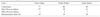
Table 3
The Concordance, Major & Minor Disconcordance and The Disconcordance Rate (%) according to Body Parts Except Wards Triangle. (F-L: Femur-L Spine, F-D: Femur-Distal Radius, L-D: Femur-Distal Radius)
![]()


References
1. Lee SJ, Suh JS, Koo JW, Ahn JC. Measurement of bone mineral density using dual energy X-ray absorptiometry in normal Korean adults. Korean J Bone Metab. 1994. 1:201–208.
2. Koh SK, Cho SH, Hwang YY, et al. Spinal bone mineral density of normal and osteoporotic women in Korea. J Korean Med Sci. 1992. 7:136–140.

3. Porgrund H, Makin M, Robin G, Menczel J, Steingerg R. Osteoporosis in patients with fractured femoral neck in Jerusaleum. Clin Orthop Relat Res. 1977. (124):165–172.
4. Riggs BL, Wakner HW, Dunn WL, Mazess RB, Offord KP, Melton LJ 3rd. Differential changes in bone mineral density of the appendicular and axial skeleton with aging: relationship to spinal osteoporosis. J Clin Invest. 1981. 67:328–335.
5. Bohr H, Schadt O. Bone mineral content of femoral bone and lumbar spine measured in women with fracture of the femoral neck by dual photon absorptiometry. Clin Orthop Relat Res. 1983. 179:240–245.
6. Bell GH, Dunber O, Beck JS, Gibb A. Variations in strength of vertebrae with age and their relation to osteoporosis. Calcif Tissue Res. 1967. 1:75–86.
7. Carter DR, Hayes WC. Bone compressive strength: the influence of density and strain rate. Science. 1976. 194:1174–1176.
8. Compston JE. Osteoporosis. Clin Endocrinol (Oxf). 1990. 33:653–682.
9. Moon WN, Oh HJ, Suh SW. Differences in bone mineral density by using different densitometers or by measuring different sites of spine in osteoporotic vertebral fractures. J Korean Orthop Assoc. 1999. 34:1153–1157.
10. Mazess RB, Barden H, Ettinger M, Schultz E. Bone density of the radius, spine and proximal femur in osteoporosis. J Bone Miner Res. 1988. 3:13–18.
11. Riggs BL, Melton LJ 3rd. Evidence for two distinct syndromes of involutional osteoporosis. Am J Med. 1983. 75:899–901.
12. Lane JM, Riely EH, Wirganowicz PZ. Osteoporosis: diagnosis and treatment. Instr Course Lect. 1997. 46:445–458.
13. Melton LJ 3rd, Wahner HW, Richelson LS, O'Fallon WM, Riggs BL. Osteoporosis and the risk of hip fracture. Am J Epidemiol. 1986. 124:254–261.
14. Bachrach LK, Hastie T, Wang MC, Narasimhan B, Marcus R. Bone mineral acquisition in health Asian, Hispanic, black, and Caucasian youth: a longitudinal study. J Clin Endocrinol Metab. 1999. 84:4702–4712.
15. Feyerabend AJ, Lear JL. Regional variations in bone mineral density as assessed with dual-energy photon absorptiometry and dual x-ray absorptiometry. Radiology. 1993. 186:467–469.
16. Choi JS, An KC, Lee CS, Choi JM, Kim JY, Shin DR. DEXA T-score concordance and discordance between hip and lumbar spine. J Korean Soc Spine Surg. 2003. 10:75–81.
17. Rowe SM, Jung ST, Lee JY. Epidemilogy of osteoporosis in Korea. Osteoporos Int. 1997. 7:Suppl 3. S88–S90.
18. Glüer CC, Steiger P, Selvidge R, Elliesen-Kliefoth K, Hayashi C, Genant HK. Comparative assessment of dual-photon absorptiometry and dual-energy radiography. Radiology. 1990. 174:223–228.
 XML Download