

 PDF
PDF ePub
ePub Citation
 Print
Print


Abstract
Purpose
We wanted to measure the femoral neck anteversion (FNA) angles using a 3D CT scan that perpendicularly cut the mechanical axis of the femur and to assess the accuracy and reproducibility of different measuring methods.
Materials and Methods
We obtained 95 cases of 3D CT images of the cross-section perpendicular to the mechanical axis of the femur. The methods used to measure the FNA angles included a method using the CT image of the area where the femoral neck is confluent to the greater trochanter (method 1), a method using the CT image taken from the neck base immediately prior to the beginning of the area of the lesser trochanter (method 2) and a method by which measurements are made after putting 3D bone models on a horizontal plane in virtual space (method 3). The reference axes of the distal femur we used were the anatomical transepicondylar axis, the surgical transepicondylar axis and the real posterior condylar axis.
Results
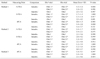
The FNA angles measured by method 1 were 4.79±6.41° to the anatomical transepicondylar axis (ATEA), 6.09±6.58° to the surgical transepicondylar axis (STEA) and 7.96±6.81° to the real posterior condylar axis (rPCA). The FNA angles measured by method 2 were 16.01±8.31° to the ATEA, 19.52±8.38° to the STEA and 21.79±8.52° to the rPCA. The FNA angles measured by method 3 were 20.15±12.89° to the rPCA.
Figures and Tables
 | Fig. 1The position of femoral head center was determined by placing a circle onto the circumference of the femoral head on a set of (A) axial and (B) coronal section views, showing the largest bone contour of the femoral head.
|
 | Fig. 2The intercondylar notch center was defined as the middle of the line connecting the narrowest anterior-to-posterior borders on an axial section.
|
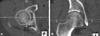
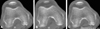
 | Fig. 3(A) Femoral neck axis of method 1 was drawn femoral neck center using of CT image on proximal-most neck confluence slice level and superimposed femoral head center. (B) Femoral neck axis of method 2 was drawn using the center of the base of the femoral neck and superimposed femoral head center.
|
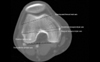 | Fig. 4Posterior condylar line is the tangent of the posterior femoral condyles, the Anatomical Trans Epicondylar Axis (TEA) connects the medial to the lateral epicondyle, the Surgical TEA connects the Medial sulcus to the lateral epicondyle.
|
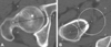
 | Fig. 5Real posterior condylar axis was drawn. (A, B) As the most lateral posterior condyle and the most medial posterior condyle did not exist on same plane, (C) real posterior condylar axis was drawn on the most medial posterior condylar plane by superimposing the most lateral posterior condyle to the medial plane. MAPC: medial apex of posterior condyle, LAPC: lateral apex of posterior condyle.
|
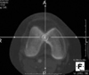
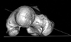 | Fig. 6Femoral neck axis of method 3 was measured on 3D bone model. On the top table position, the 3D bone model aligned to the mechanical axis. femoral neck axis was defined by the line drawn using femoral head center and the center of the middle of the line connecting the narrowest anterosuperior and posteroinferior border of the neck.
|


References
1. Kingsley PC, Olmsted KL. A study to determine the angle of anteversion of the neck of the femur. J Bone Joint Surg Am. 1948. 30A:745–751.

2. Kuo TY, Skedros JG, Bloebaum RD. Measurement of femoral anteversion by biplane radiography and computed tomography imaging: comparison with an anatomic reference. Invest Radiol. 2003. 38:221–229.
3. Billing L. Roentgen examination of the proximal femur end in children and adolescents; a standardized technique also suitable for determination of the collum-, anteversion-, and epiphyseal angles; a study of slipped epiphysis and coxa plana. Acta Radiol Suppl. 1954. 110:1–80.
4. Weiner DS, Cook AJ, Hoyt WA Jr, Oravec CE. Computed tomography in the measurement of femoral anteversion. Orthopedics. 1978. 1:299–306.
5. Reikerås O, Bjerkreim I, Kolbenstvedt A. Anteversion of the acetabulum and femoral neck in normals and in patients with osteoarthritis of the hip. Acta Orthop Scand. 1983. 54:18–23.
6. Murphy SB, Simon SR, Kijewski PK, Wilkinson RH, Griscom NT. Femoral anteversion. J Bone Joint Surg Am. 1987. 69:1169–1176.
7. Sugano N, Noble PC, Kamaric E. A comparison of alternative methods of measuring femoral anteversion. J Comput Assist Tomogr. 1998. 22:610–614.
8. Lee YS, Oh SH, Seon JK, Song EK, Yoon TR. 3D femoral neck anteversion measurements based on the posterior femoral plane in ORTHODOC system. Med Biol Eng Comput. 2006. 44:895–906.
9. Kim JS, Park TS, Park SB, Kim JS, Kim IY, Kim SI. Measurement of femoral neck anteversion in 3D. Part 1: 3D imaging method. Med Biol Eng Comput. 2000. 38:603–609.
10. Kim JS, Park TS, Park SB, Kim JS, Kim IY, Kim SI. Measurement of femoral neck anteversion in 3D. Part 2: 3D modelling method. Med Biol Eng Comput. 2000. 38:610–616.
11. Hermann KL, Egund N. CT measurement of anteversion in the femoral neck. The influence of femur positioning. Acta Radiol. 1997. 38:527–532.
12. Hernandez RJ, Tachdjian MO, Poznanski AK, Dias LS. CT determination of femoral torsion. AJR Am J Roentgenol. 1981. 137:97–101.
13. Dunn DM. Anteversion of the neck of the femur; a method of measurement. J Bone Joint Surg Br. 1952. 34-B:181–186.
14. Edholm P. Nomogram for measuring the anteversion angle and angulation of fracture from roentgenograms. Acta Radiol Diagn (Stockh). 1972. 12:856–864.
15. Ryder CT, Crane L. Measuring femoral anteversion; the problem and a method. J Bone Joint Surg Am. 1953. 35-A:321–328.
16. Henriksson L. Measurement of femoral neck anteversion and inclination. A radiographic study in children. Acta Orthop Scand Suppl. 1980. 186:1–59.
17. Backman S. The proximal end of the femur: investigations with special reference to the etiology of femoral neck fractures; anatomical studies; roentgen projections; theoretical stress calculations; experimental production of fractures. Acta Radiol Suppl. 1957. 146:1–166.
18. Griffin FM, Insall JN, Scuderi GR. The posterior condylar angle in osteoarthritic knees. J Arthroplasty. 1998. 13:812–815.
19. Newbern DG, Faris PM, Ritter MA, Keating EM, Meding JB, Berend ME. A clinical comparison of patellar tracking using the transepicondylar axis and the posterior condylar axis. J Arthroplasty. 2006. 21:1141–1146.
 XML Download