

 PDF
PDF ePub
ePub Citation
 Print
Print


Abstract
Purpose
We propose to improve the use of Proximal Femoral Nail Anti-rotation in Korea by reporting anatomical measurements of the normal Korean proximal femur.
Materials and Methods
A total of 230 patients were enrolled who had all experienced a femoral intertrochanteric fracture and undergone the Proximal Femoral Nail Anti-rotation surgical procedure between February 2007 and April 2011. We measured the neck-shaft angle and endosteal width at the isthmus of a normal femur, and the distance between the greater trochanter and the nail tip of the Proximal Femoral Nail Anti-rotation in post-operative plain radiography. We analyzed the average and standard deviations of the measurements. We also investigated correlations with the patient gender.
Results
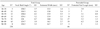
The average neck-shaft angle and endosteal width at the isthmus were 129.7°(111.0~138.3°) and 14.5 mm (9.7~23.1 mm), respectively. The average protruded nail length was 4.9 mm (1.0~15.0 mm). The femur neck-shaft angle had a correlation ratio with gender (p=0.000). However, the endosteal width at the isthmus level and the protruded nail length had no correlation ratio with gender (p=0.108, 0.573, respectively).
Conclusion
Until now, in intertrochanteric fracture operations using Proximal Femoral Nail Anti-rotation, the selection of devices has been extremely limited. Through this study we present the average Korean anatomical neck-shaft angle, endosteal width of the femur, and protruding length of the nail tip. We expect that these numerical values can be used in the production of new devices with shorter proximal nails, which would be more appropriates for Koreans.
Figures and Tables
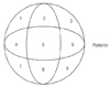 | Fig. 1For the Cleveland Index. The femoral head (axial view) was devided into nine zone to document the position of the tip of the blade.
|
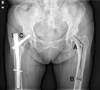 | Fig. 2Pelvis AP X-ray imaging: The method of measuring (A) the neck-shaft angle, (B) endosteal width at the isthmus level, (C) protruded nail tip length.
|
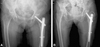 | Fig. 3Comparison 2 cases which use same size (10 mm diameter, 130° angle): (A) Contra-lateral neck-shaft angle was 131.1° and protruded nail tip length was checked 1 mm. (B) Contra-lateral neck-shaft angle was 136.2° and protruded nail tip length was checked 9.2 mm.
|
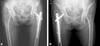 | Fig. 4Comparison 2 cases which has same neck-shaft angle (about 135°): (A) Using of 130°-angulated PFNA and protruded nail length was checked 0 mm. (B) Using of 125°-angulated PFNA and protruded nail length was checked 5.6 mm.
|
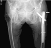
Table 1
Mean Values and P-Values of Variables Measured between the Male and the Female Subjects (N=230)

![]()

References
1. Hwang DS, Rhee KJ, Choi JH. Recovery of walking ability after treatment of unstable intertrochanteric fractures in elderly patients: comparison of compression hip screw to primary hemiarthroplasty. J Korean Hip Soc. 1999. 11:22–29.
2. Lee JY, Lee SY. Treatment of proximal femoral extracapsular fracture with proximal femoral nail antirotation (PFNA): comparison with proximal femoral nail (PFN). J Korean Hip Soc. 2007. 19:183–189.

3. Richmond J, Aharonoff GB, Zuckernab JD, Koval KJ. Mortality risk after hip fracture. J Orthop Trauma. 2003. 17:S2–S5.
4. Hornby R, Evans JG, Vardon V. Operative or conservative treatment for trochanteric fractures of the femur: A randomised epidemiological trial in elderly patients. J Bone Joint Surg Br. 1989. 71:619–623.
5. Jensen JS. Determining factors for the mortality following hip fractures. Injury. 1984. 15:411–414.
6. Kyle RF, Cabanela ME, Russel TA, et al. Fractures of the proximal part of the femur. Instr Course Lect. 1995. 44:227–253.
7. Pajarinen J, Lindahl J, Michelsson O, Savolainen V, Hirvensalo E. Peritrochanteric femoral fractures treated with a dymamic hip screw or a proximal femoral nail. A randomised study comparing post-operative rehabilitation. J Bone Joint Surg Br. 2005. 87:76–81.
8. Chang SA, Cho YH, Byun YS, Han JH, Park JY, Lee CY. The treatment of trochanteric femoral fracture with using proximal femoral nail antirotation (PFNA). J Korean Hip Soc. 2009. 21:252–256.
9. Park MS, Lim YJ, Kim YS, Kim KH, Cho HM. Treatment of the proximal femoral fractures with proximal femoral nail antirotation (PFNA). J Korean Fract Soc. 2009. 22:91–97.
10. Lee KB, LEE BT. Complications of femoral peritrochanteric fractures treated with proximal femoral nail (PFN). J Korean Fract Soc. 2007. 20:33–39.
11. Cleveland M, Bosworth DM, Thompson FR, Wilson HJ Jr, Ishizuka T. A ten-year analysis of intertrochanteric fractures of the femur. J Bone Joint Surg Am. 1959. 41-A:1399–1408.
12. Chung YK, Hwang JH, Kim HK. The treatment of peritrochanteric fracture of femur with proximal femoral nail: Comparative study with dynamic hip screw. J Korean Hip Soc. 2007. 19:167–175.
13. Hardy DC, Descamps PY, Krallis P, et al. Use of an intramedullary hip-screw compared with a compression hip-screw with a plate for intertrochanteric femoral fractures. A prospective, randomized study of one hundred patients. J Bone Joint Surg Am. 1998. 80:618–630.
14. Strauss E, Frank J, Lee J, Kummer FJ, Tejwani N. Helical blade versus sliding hip screw for treatment of unstable intertrochanteric hip fractures: a biomechanical evaluation. Injury. 2006. 37:984–989.
15. Suh KT, Lee SH, Cho BM. Radiologic analysis of the proximal femoral morphology in normal Korean adults. J Korean Orthop Assoc. 1999. 34:891–897.
16. Khang G, Choi KW, Kim CS, Yang JS, Bae TS. A study of Korean femoral geometry. Clin Orthop Relat Res. 2003. 406:116–122.
17. Pajarinen J, Lindahl J, Michelsson O, Savolainen V, Hirvensalo E. The morphology of the proximal femur. J Bone Joint Surg Br. 1992. 74:28–32.
 XML Download