

 PDF
PDF ePub
ePub Citation
 Print
Print


Abstract
Multiple drilling of the femoral head is a commonly-used surgical technique for the treatment of osteonecrosis. Several holes are drilled into the femoral head using a small-diameter Steinmann pin. This decompression technique promotes revascularization and regeneration of necrotic tissue in the femoral head. Known complications are heterotopic ossification and femoral subtrochanteric fractures, although these have only been rarely reported. Our case study focuses on a subtrochanteric fracture 6 weeks after a multiple drilling operation.
Figures and Tables
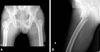
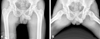 | Fig. 1Anteroposterior and frog-lateral view of both hip radiographs show sclerotic change of right femoral head.
|
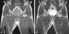 | Fig. 2(A) Coronal T1-weighted (SE; TR629/TE10 msec) image shows decreased signal at subchondral location. Also it shows osteonecrotic segment separated from normal bone by low signal intensity. (B) Coronal T2-weighted image shows increased signal at subchondral location of necrotic tissue.
|
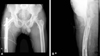
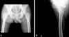 | Fig. 3Immediate postoperative anteroposterior and lateral radiographs of the right hip show multiple radiolucent lines those from lateral cortex of proximal femur to femoral head.
|
 | Fig. 4Plain radiograph after 6 weeks of operation shows multiple lines without any bone destruction or fracture.
|
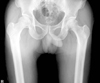
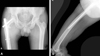
 | Fig. 5(A) Anteroposterior radiograph of right hip shows transverse fracture line at subtrochanteric area and it started just inferior of drilling site. (B) Lateral radiograph of right hip shows complete fracture line and posterior displacement of distal fragment of proximal femur.
|
 | Fig. 6Immediate postoperative anteroposterior and lateral radiographs of the right hip show intramedullary fixation status using PFNA II (Synthes, Oberdorf, Swiss).
|
References
1. Marker DR, Seyler TM, McGrath MS, Delanois RE, Ulrich SD, Mont MA. Treatment of early stage osteonecrosis of the femoral head. J Bone Joint Surg Am. 2008. 90:Suppl 4. 175–187.

2. Kim YM, Lee DY, Chung MS, Kang SB, Kim BS, Kim HJ. Result of multiple drilling for the early stage nontraumatic osteonecrosis of the femoral head. J Korean Orthop Assoc. 1997. 32:977–983.
3. Kim SY, Kim DH. The results of multiple drilling for the treatment of osteonecrosis of the femoral head. J Korean Hip Soc. 1999. 11:30–38.
4. Mont MA, Ragland PS, Etienne G. Core decompression of the femoral head for osteonecrosis using percutaneous multiple small-diameter drilling. Clin Orthop Relat Res. 2004. 429:131–138.
5. Marker DR, Seyler TM, Ulrich SD, Srivastava S, Mont MA. Do modern techniques improve core decompression outcomes for hip osteonecrosis? Clin Orthop Relat Res. 2008. 466:1093–1103.
6. Song WS, Yoo JJ, Kim YM, Kim HJ. Results of multiple drilling compared with those of conventional methods of core decompression. Clin Orthop Relat Res. 2007. 454:139–146.
7. Colwell CW Jr. The controversy of core decompression of the femoral head for osteonecrosis. Arthritis Rheum. 1989. 32:797–800.
8. Berend KR, Gunneson EE, Urbaniak JR. Free vascularized fibular grafting for the treatment of postcollapse osteonecrosis of the femoral head. J Bone Joint Surg Am. 2003. 85-A:987–993.
9. Fung DA, Frey S, Menkowitz M, Mark A. Subtrochanteric fracture in a patient with trabecular metal osteonecrosis intervention implant. Orthopedics. 2008. 31:614.
10. Lieberman JR, Berry DJ, Mont MA, et al. Osteonecrosis of the hip: management in the 21st century. Instr Course Lect. 2003. 52:337–355.
 XML Download