

 PDF
PDF ePub
ePub Citation
 Print
Print


Abstract
Purpose
We evaluated the usefulness of radiographic parameters for osteoporosis by analyzing the results of radiographic parameters determined by digital hip radiographs and bone mineral density T-scores, as assessed by Dual Energy X-ray Absorptiometry (DEXA).
Materials and Methods
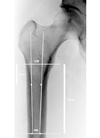
The authors reviewed 100 subjects in the hip fracture group and 50 in the non-fracture control group. Digital hip radiographs were assessed to determine the values of Singh index, Canal-to-Calcar Ratio, and Cortical Thickness Index (CTI). Bone mineral density was assessed by DEXA.
Results
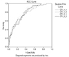
Intraclass Correlation Coefficient (ICC) results of the CTI were above 0.8 in the fracture group. Compared to the control group, the fracture group showed higher ICCs. Interobserver ICCs were especially lower in the control group. There were statistically significant correlations between CTI and DEXA (r=0.50~0.58, p<0.05). In the analysis of ROC curves, a mean threshold for CTI set a value of 0.54 (0.53~0.55), and mean sensitivity and specificity were 75.5% (69~79%) and 67.8% (65~78%), respectively.
Figures and Tables
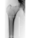 | Fig. 2Cortical thickness index measurement on anteroposterior radiograph of the hip. Cortical thickness index is equal to femoral diaphysis with minus intramedullary width divided by diaphysis width.
|
References
1. Aloia JF, Vaswani A, Atkins H, Zanzi I, Ellis K, Cohn SH. Radiographic morphometry and osteopenia in spinal osteoporosis. J Nucl Med. 1977. 18:425–431.
2. Devlin H, Karayianni K, Mitsea A, et al. Diagnosing osteoporosis by using dental panoramic radiographs: the OSTEODENT project. Oral Surg Oral Med Oral Pathol Oral Radiol Endod. 2007. 104:821–828.

3. Singh M, Nagrath AR, Maini PS. Changes in trabecular pattern of the upper end of the femur as an index of osteoporosis. J Bone Joint Surg Am. 1970. 52:457–467.
4. Sah AP, Thornhill TS, Leboff MS, Glowacki J. Correlation of plain radiographic indices of the hip with quantitative bone mineral density. Osteoporos Int. 2007. 18:1119–1126.
5. Dorr LD, Faugere MC, Mackel AM, Gruen TA, Bognar B, Malluche HH. Structural and cellular assessment of bone quality of proximal femur. Bone. 1993. 14:231–242.
6. Boehm HF, Link TM. Bone imaging: traditional techniques and their interpretation. Curr Osteoporos Rep. 2004. 2:41–46.
7. O'Gradaigh D, Debiram I, Love S, Richards HK, Compston JE. A prospective study of discordance in diagnosis of osteoporosis using spine and proximal femur bone densitometry. Osteoporos Int. 2003. 14:13–18.
8. Astrand J, Thorngren KG, Tägil M, Akesson K. 3-year follow-up of 215 fracture patients from a prospective and consecutive osteoporosis screening program. Fracture patients care! Acta Orthop. 2008. 79:404–409.
9. Bergmann P, Body JJ, Boonen S, et al. Evidence-based guidelines for the use of biochemical markers of bone turnover in the selection and monitoring of bisphosphonate treatment in osteoporosis: a consensus document of the Belgian Bone Club. Int J Clin Pract. 2009. 63:19–26.
10. Civitelli R, Armamento-Villareal R, Napoli N. Bone turnover markers: understanding their value in clinical trials and clinical practice. Osteoporos Int. 2009. 20:843–851.
11. Szulc P, Delmas PD. Biochemical markers of bone turnover: potential use in the investigation and management of postmenopausal osteoporosis. Osteoporos Int. 2008. 19:1683–1704.
12. Kawashima T, Uhthoff HK. Pattern of bone loss of the proximal femur: a radiologic, densitometric, and histomorphometric study. J Orthop Res. 1991. 9:634–640.
13. Koot VC, Kesselaer SM, Clevers GJ, de Hooge P, Weits T, van der Werken C. Evaluation of the Singh index for measuring osteoporosis. J Bone Joint Surg Br. 1996. 78:831–834.
14. Pogrund H, Rigal WM, Makin M, Robin G, Menczel J, Steinberg R. Determination of osteoporosis in patients with fractured femoral neck using the Singh index: a Jerusalem study. Clin Orthop Relat Res. 1981. 156:189–195.
15. Lewiecki EM, Baim S, Langman CB, Bilezikian JP. The official positions of the International Society for Clinical Densitometry: perceptions and commentary. J Clin Densitom. 2009. 12:267–271.
16. Watts NB. Fundamentals and pitfalls of bone densitometry using dual-energy X-ray absorptiometry (DXA). Osteoporos Int. 2004. 15:847–854.
17. Masud T, Jawed S, Doyle DV, Spector TD. A population study of the screening potential of assessment of trabecular pattern of the femoral neck (Singh index): the Chingford Study. Br J Radiol. 1995. 68:389–393.
18. Miller RG. Osteoporosis in postmenopausal women. Therapy options across a wide range of risk for fracture. Geriatrics. 2006. 61:24–30.
19. Hauschild O, Ghanem N, Oberst M, et al. Evaluation of Singh index for assessment of osteoporosis using digital radiography. Eur J Radiol. 2009. 71:152–158.
20. Landis JR, Koch GG. The measurement of observer agreement for categorical data. Biometrics. 1977. 33:159–174.
21. Wagner S, Stäbler A, Sittek H, et al. Diagnosis of osteoporosis: visual assessment on conventional versus digital radiographs. Osteoporos Int. 2005. 16:1815–1822.
22. Lindhardt FE. Clinical experiences with computed radiography. Eur J Radiol. 1996. 22:175–185.
23. Okamura T, Tanaka S, Koyama K, et al. Clinical evaluation of digital radiography based on a large-area cesium iodide-amorphous silicon flat-panel detector compared with screen-film radiography for skeletal system and abdomen. Eur Radiol. 2002. 12:1741–1747.
 XML Download