

 PDF
PDF ePub
ePub Citation
 Print
Print


Abstract
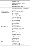
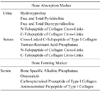
Osteoporosis is defined as a skeletal disorder characterized by compromised bone strength, predisposing an individual to increased fracture risk. Many factors can lead to the development of osteoporosis. It is usually asymptomatic unless osteoporotic fracture and secondary changes of bone structure occur. Early radiographs show normal findings; however, osteopenic appearance, fracture, cortical bone thinning, and roughening of bone trabeculae can be found according to severity of osteoporosis. These symptoms are most frequently found in the spine and proximal femur. Bone mineral density (BMD) is the standard method used to diagnose osteoporosis, and dual energy X-ray absorptiometry (DXA), one of the measurement tools for BMD, is particularly regarded as the appropriate tool applicable to WHO criteria, which defines osteoporosis as a T-score of less than 2.5 SDs below the mean of young adult women. Peripheral densitometry is less useful in predicting the risk of fractures of the spine and proximal femur, and it is not enough to diagnose and treat osteoporosis. Biochemical bone markers have demonstrated utility in clinical research and trials; however, they cannot replace BMD as a diagnostic tool. WHO recently developed FRAX, a novel method we can use to more conveniently evaluate osteoporotic fracture risk.
Figures and Tables
References
1. Consensus development conference: diagnosis, prophylaxis, and treatment of osteoporosis. Am J Med. 1993. 94:646–650.
2. National Institute of Health. Osteoporosis prevention, diagnosis, and therapy. NIH Consens Statement. 2000. 17:1–45.
3. Lane NE. Epidemiology, etiology, and diagnosis of osteoporosis. Am J Obstet Gynecol. 2006. 194:2 Suppl. S3–S11.

4. Lee KH. Diagnosis of Osteoporosis. J Korean Hip Soc. 2007. 19:260–265.
5. Kim N, Rowe BH, Raymond G, et al. Underreporting of vertebral fractures on routine chest radiography. AJR Am J Roentgenol. 2004. 182:297–300.
6. Cooper C, Atkinson EJ, O'Fallon WM, Melton LJ 3rd. Incidence of clinically diagnosed vertebral fractures: a population-based study in Rochester, Minnesota, 1985-1989. J Bone Miner Res. 1992. 7:221–227.
7. Mayo-Smith W, Rosenthal DI. Radiographic appearance of osteopenia. Radiol Clin North Am. 1991. 29:37–47.
8. Johnston CC Jr, Epstein S. Clinical, biochemical, radiographic, epidemiologic, and economic features of osteoporosis. Orthop Clin North Am. 1981. 12:559–569.
9. Yang SO, Kim S, Juhng SK. Imaging diagnosis of osteoporotic fracture. J Korean Med Assoc. 2010. 53:67–75.
10. Haller J, André MP, Resnick D, et al. Detection of thoracolumbar vertebral body destruction with lateral spine radiography. Part I: Investigation in cadavers. Invest Radiol. 1990. 25:517–522.
11. Singh M, Nagrath AR, Maini PS. Changes in trabecular pattern of the upper end of the femur as an index of osteoporosis. J Bone Joint Surg Am. 1970. 52:457–467.
12. Cummings SR, Kelsey JL, Nevitt MC, O'Dowd KJ. Epidemiology of osteoporosis and osteoporotic fractures. Epidemiol Rev. 1985. 7:178–208.
13. Neff MJ. ACOG releases guidelines for clinical management of osteoporosis. Am Fam Physician. 2004. 69:1558. 1560.
14. The Korean Society of Bone Metabolism. Physician's guideline for osteoporosis. 2007. Seoul: Seoheung Publishing Co.;21–33.
15. Pouilles JM, Tremollieres F, Ribot C. Spine and femur densitometry at the menopause: are both sites necessary in the assessment of the risk of osteoporosis? Calcif Tissue Int. 1993. 52:344–347.
16. Leib ES, Lewiecki EM, Binkley N, Hamdy RC. Official positions of the International Society for Clinical Densitometry. J Clin Densitom. 2004. 7:1–6.
17. Blake GM, Fogelman I. Bone densitometry and the diagnosis of osteoporosis. Semin Nucl Med. 2001. 31:69–81.
18. Patel R, Blake GM, Rymer J, Fogelman I. Long-term precision of DXA scanning assessed over seven years in forty postmenopausal women. Osteoporos Int. 2000. 11:68–75.
19. Ravn P, Overgaard K, Huang C, Ross PD, Green D, McClung M. Comparison of bone densitometry of the phalanges, distal forearm and axial skeleton in early postmenopausal women participating in the EPIC Study. Osteoporos Int. 1996. 6:308–313.
20. Kullenberg R, Falch JA. Prevalence of osteoporosis using bone mineral measurements at the calcaneus by dual X-ray and laser (DXL). Osteoporos Int. 2003. 14:823–827.
21. Committee on Gynecologic Practice. Bone density screening for osteoporosis. Int J Gynaecol Obstet. 2002. 77:299–301.
22. Gonnelli S, Cepollaro C, Montagnani A, et al. Heel ultrasonography in monitoring alendronate therapy: a four-year longitudinal study. Osteoporos Int. 2002. 13:415–421.
23. Murray JF. Primer on the metabolic bone diseases and disorders of mineral metabolism. 2003. 5th ed. Washington DC: American Society for Bone and Mineral Research;307–360.
24. Blake GM, Fogelman I. Peripheral or central densitometry: does it matter which technique we use? J Clin Densitom. 2001. 4:83–96.
25. The Korean Society of Bone Metabolism. Osteoporosis. 2006. 3rd ed. Seoul: Hanmi-Euihak;113–181.
26. Kanis JA. WHO Study Group. Assessment of fracture risk and its application to screening for postmenopausal osteoporosis: synopsis of a WHO report. Osteoporos Int. 1994. 4:368–381.
27. Kanis JA, Oden A, Johansson H, Borgström F, Ström O, McCloskey E. FRAX and its applications to clinical practice. Bone. 2009. 44:734–743.
28. Kanis JA, Johnell O, Oden A, Johansson H, McCloskey E. FRAX and the assessment of fracture probability in men and women from the UK. Osteoporos Int. 2008. 19:385–397.
 XML Download