

 PDF
PDF ePub
ePub Citation
 Print
Print


Abstract
Purpose
For inserting an acetabular cup with the correct inclination angle, we checked the pelvic tilts using the lateral decubitus position X-ray and a goniometer and pointer. The accuracy of the cup inclination at the targeted angles was evaluated after insertion of the cup at an adjusted angle with using a goniometer and pointer.
Materials and Methods
Between January 2008 and December 2009, 56 hips in 50 patients who underwent total hip replacement arthroplasty (THRA) were enrolled. The mean age at the time of surgery was 63.8 years. There were 31 male patients (36 hips) and 19 female patients (20 hips). The preoperative diagnoses included osteonecrosis of the femoral head in 27 hips, secondary osteoarthritis in 10 hips and femoral neck fracture in 14 hips. The preoperative pelvic tilts were evaluated according to the lateral decubitus position X-ray with using a goniometer and pointer. The target inclination angle was 40° for 27 hips that underwent ceramic-on-ceramic THRA. The target inclination angle was 45° for the 29 hips that underwent ceramic-on-polyethylene THRA. The inclination of the cup was evaluated after inserting the acetabular cup at the adjusted angle using a goniometer and pointer.
Figures and Tables
 | Fig. 1(A) Goniometer with long pointer targeted to 45° for measuring the pelvic tilt. (B) Goniometer and pointer can be divided for sterilization.
|
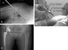 | Fig. 2(A) Preoperative AP X-ray with goniometer and pointer. An angle between inter-teardrop line and pointer is 47.3°. Pelvis was tilted to cephalad 2.3°. (B) The photograph of acetabular cup insertion using goniometer and pointer. This hip was planned for ceramic on ceramic THA(target angle was 40°), so goniometer was fixed at 42.3°. Cup fixation was done parallel to goniometer and pointer. (C) Post-operative inclination angle of cup was 40.4°.
|


References
1. Ali Khan MA, Brakenbury PH, Reynolds IS. Dislocation following total hip replacement. J Bone Joint Surg Br. 1981. 63-B:214–218.

2. Engh CA, Massin P, Suthers KE. Roentgenographic assessment of the biologic fixation of porous-surfaced femoral components. Clin Orthop Relat Res. 1990. 257:107–128.
3. Lewinnek GE, Lewis JL, Tarr R, Compere CL, Zimmerman JR. Dislocations after total hip-replacement arthroplasties. J Bone Joint Surg Am. 1978. 60:217–220.
4. D'Lima DD, Urquhart AG, Buehler KO, Walker RH, Colwell CW Jr. The effect of the orientation of the acetabular and femoral components on the range of motion of the hip at different head-neck ratios. J Bone Joint Surg Am. 2000. 82:315–321.
5. Del Schutte H Jr, Lipman AJ, Bannar SM, Livermore JT, Ilstrup D, Morrey BF. Effects of acetabular abduction on cup wear rates in total hip arthroplasty. J Arthroplasty. 1998. 13:621–626.
6. Kennedy JG, Rogers WB, Soffe KE, Sullivan RJ, Griffen DG, Sheehan LJ. Effect of acetabular component orientation on recurrent dislocation, pelvic osteolysis, polyethylene wear, and component migration. J Arthroplasty. 1998. 13:530–534.
7. Lucas DH, Scott RD. The Ranawat sign. A specific maneuver to assess component positioning in total hip arthroplasty. J Orthop Tech. 1994. 2:59–61.
8. Wolf A, Digioia AM 3rd, Mor AB, Jaramaz B. Cup alignment error model for total hip arthroplasty. Clin Orthop Relat Res. 2005. 437:132–137.
9. Hedlundh U, Ahnfelt L, Hybbinette CH, Weckstrom J, Fredin H. Surgical experience related to dislocations after total hip arthroplasty. J Bone Joint Surg Br. 1996. 78:206–209.
10. Ha YC, Kim SY, Kim HJ, Yoo JJ, Koo KH. Ceramic liner fracture after cementless alumina-on-alumina total hip arthroplasty. Clin Orthop Relat Res. 2007. 458:106–110.
11. Park YS, Hwang SK, Choy WS, Kim YS, Moon YW, Lim SJ. Ceramic failure after total hip arthroplasty with an alumina-on-alumina bearing. J Bone Joint Surg Am. 2006. 88:780–787.
12. Han KY, Cho SG. Radiologic examination at lateral decubitus position for reducing the variability of cup inclination. J Korean Orthop Assoc. 2009. 44:408–413.
13. Crowninshield RD, Maloney WJ, Wentz DH, Humphrey SM, Blanchard CR. Biomechanics of large femoral heads: what they do and don't do. Clin Orthop Relat Res. 2004. 429:102–107.
14. Daly PJ, Morrey BF. Operative correction of an unstable total hip arthroplasty. J Bone Joint Surg Am. 1992. 74:1334–1343.
15. DiGioia AM, Jaramaz B, Blackwell M, et al. The Otto Aufranc Award. Image guided navigation system to measure intraoperatively acetabular implant alignment. Clin Orthop Relat Res. 1998. 355:8–22.
16. Digioia AM 3rd, Jaramaz B, Plakseychuk AY, et al. Comparison of a mechanical acetabular alignment guide with computer placement of the socket. J Arthroplasty. 2002. 17:359–364.
17. Belei P, Skwara A, De La Fuente M, et al. Fluoroscopic navigation system for hip surface replacement. Comput Aided Surg. 2007. 12:160–167.
18. Petrella AJ, Stowe JQ, D'Lima DD, Rullkoetter PJ, Laz PJ. Computer-assisted versus manual alignment in THA: a probabilistic approach to range of motion. Clin Orthop Relat Res. 2009. 467:50–55.
19. Thorey F, Klages P, Lerch M, Flörkemeier T, Windhagen H, von Lewinski G. Cup positioning in primary total hip arthroplasty using an imageless navigation device: is there a learning curve? Orthopedics. 2009. 32:10 Suppl. 14–17.
20. Hassan DM, Johnston GH, Dust WN, Watson G, Dolovich AT. Accuracy of intraoperative assessment of acetabular prosthesis placement. J Arthroplasty. 1998. 13:80–84.
21. Park SE, Kim WY, Kwon OS, Kim YY, Won HY. Intraoperative imaging to monitor the alignment of acetabular cup for total hip replacement arthroplasty. J Korean Hip Soc. 2007. 19:155–160.
22. Nizard RS, Sedel L, Christel P, Meunier A, Soudry M, Witvoet J. Ten-year survivorship of cemented ceramic-ceramic total hip prosthesis. Clin Orthop Relat Res. 1992. 282:53–63.
23. Garino JP. Modern ceramic-on-ceramic total hip systems in the United States: early results. Clin Orthop Relat Res. 2000. 379:41–47.
24. Bosker BH, Verheyen CC, Horstmann WG, Tulp NJ. Poor accuracy of freehand cup positioning during total hip arthroplasty. Arch Orthop Trauma Surg. 2007. 127:375–379.
25. Saxler G, Marx A, Vandevelde D, et al. The accuracy of free-hand cup positioning--a CT based measurement of cup placement in 105 total hip arthroplasties. Int Orthop. 2004. 28:198–201.
26. Kim YL, Shin SI, Nam KW, Yoo JJ, Kim YM, Kim HJ. Total hip arthroplasty for bilaterally ankylosed hips. J Arthroplasty. 2007. 22:1037–1041.
 XML Download