

 PDF
PDF ePub
ePub Citation
 Print
Print


Introduction
Both repetitive electrical and magnetic stimulations can either potentiate or decrease cortical excitability depending on the stimulation conditions,1 potentially making it useful in treating many neurological conditions such as depression, tremor, and epilepsy. Both high- and low-frequency repetitive transcranial magnetic stimulation (rTMS) of the prefrontal cortex has shown beneficial effects in patients with depression.2,3 The therapeutic effects of low-frequency rTMS (from 0.33 to 1 Hz) of the vertex or seizure foci have been investigated in epilepsy patients.4,5 Low-frequency (1-Hz) rTMS is known to decrease cortical excitability in the normal human ipsilateral motor or premotor cortex,6,7 suggesting short-term inhibition of the stimulated cortex. However, the optimal stimulation paradigm and exact therapeutic mechanism in each clinical condition is not completely understood. Given that the therapeutic efficacy needs to be maintained, repeated sessions of rTMS could be more helpful in these clinical conditions. Also, the feasibility, safety, and detailed neurophysiologic changes associated with repeated rTMS sessions have not been investigated.
There is increasing evidence that temporal cortex stimulation is helpful in certain clinical conditions, especially tinnitus and temporal lobe epilepsy.4,8 Our previous study found that 1-Hz rTMS of the temporal cortex enhanced the ipsilateral dominant cortico-cortical interaction by interhemispheric asymmetric coupling from the stimulated cortical area.9 However, functional changes in cortical and subcortical networks after 1-Hz rTMS of the temporal cortex are not completely understood.
Functional brain imaging has been undertaken to answer some of these questions. The use of positron emission tomography (PET) and single photon emission computed tomography to investigate the effects of high- and low-frequency rTMS has revealed changes in brain activity and associated effects in motor and distant cortical areas.10-12 The application of high-frequency (5-Hz) rTMS to the primary motor cortex induces a region-specific increase in the resting regional cerebral blood flow (rCBF),12 while 1-Hz rTMS applied to the same area reportedly increases rCBF locally in an intensity-dependent manner.11 These studies found that rCBF was reduced in other cortical regions, especially the medial frontal and occipital areas. Other studies have found that rTMS induces changes in rCBF and glucose metabolism in the bilateral motor and supplementary motor areas.10,13
Especially, application of 2-Hz rTMS to the primary sensorimotor cortex activated the frontal premotor and stimulated areas.10 The above-mentioned studies addressed short-term effects, while another study that assessed the long-term effects of rTMS in monkeys found that 5-Hz rTMS applied to the precentral gyrus reduces glucose metabolism in the motor and premotor cortices and increased metabolism in the cingulate and orbitofrontal cortices for a period lasting at least 8 days.14 One recent study demonstrated decreased functional magnetic resonance imaging activation in pain networks including the secondary somatosensory cortex after 10-Hz rTMS of the primary motor cortex.15 However, the effects of rTMS on local and global cortical reorganization and the precise underlying mechanisms are still unknown.
This study investigated changes in cortical activities both in and remote from the stimulated area after administering focal 1-Hz rTMS to the temporal cortex using measures of transcranial magnetic stimulation (TMS) indices and the fluorodeoxyglucose (FDG)-PET glucose metabolism. We also evaluated the feasibility, safety, and underlying neurophysiologic changes after repeated rTMS sessions in normal healthy volunteers, which is necessary before clinical trials involving large numbers of patients can be conducted.
Methods
Subjects
The subjects who participated in this study were all healthy volunteers aged between 20 to 35 years with no history of neurological disorders or head injuries. Twenty-two subjects (active rTMS group) received rTMS (age 24.4±6.4 years old, 11 males and 11 females), while 5 (sham group) received sham stimulation (24.0±4.8 years old, 2 males and 3 females). Informed consent was obtained from all subjects after the purpose and possible consequences of the study had been explained to them, according to the protocol approved by the Human Investigation Committee of our institution and the ethical principles of the Declaration of Helsinki. Patients were carefully evaluated and asked about any discomforts or concerns they had about the experiment before, during, and after each rTMS session.
TMS
All subjects underwent daily rTMS or sham rTMS for 5 consecutive days, and motor cortical excitability was measured prior to and after daily rTMS sessions and at 2 weeks after the last stimulation session (Fig. 1).
For single and repetitive pulses, TMS was delivered through a focal figure-of-eight magnetic coil (with an external diameter of 15 cm) connected to a magnetic stimulator (MagPro Rapid Rate Magnetic Stimulator, Medtronic, Shoreview, MN, USA).
Indices of motor cortical excitability
TMS indices of motor cortical excitability were measured by single- or paired-pulse TMS using surface EMG Ag-AgCl electrodes placed over the first dorsal interosseous (FDI) muscle in a belly-tendon montage. EMG raw signals were amplified, bandpass filtered (5 Hz to 5 kHz), and recorded on a personal computer using data collection and averaging software (Toennies Neuroscreen Plus, Hoechberg, Germany). TMS was delivered through the above-mentioned figure-of-eight magnetic coil from a magnetic stimulator. Subjects were seated in an armchair. The intersection of two wings of the coil was placed tangentially to the scalp with the handle pointing backward and laterally at 45° from the midlines in order to optimally activate corticospinal pathways.16 The TMS coil was placed flat on the scalp at the optimal positions to activate contralateral FDI muscles. The parameters described below were measured in all subjects to determine the motor cortical excitability before and after daily 1-Hz rTMS or sham rTMS sessions, and 2 weeks after the last stimulation session.
Resting motor threshold
The resting motor threshold (RMT) was defined as the minimum stimulus intensity required to induce a motor evoked potential (MEP) with a peak-to-peak amplitude of >50 µV in at least five of ten consecutive trials in the contralateral FDI.17 Stimulus intensities were changed in steps of 1%.
Recruitment curve
The recruitment curve (RC) to TMS was determined at intensities of 100%, 120%, and 140% RMT18,19 in each subject. Ten consecutive MEPs were recorded at each stimulus intensity. Peak-to-peak amplitudes were measured during each trial and later averaged to characterize the amplitude at each stimulus intensity. The normalized MEP was expressed as the absolute MEP amplitude relative to the maximal peripheral M wave evoked by stimulating the ulnar nerve at the wrist.
Cortical silent period
The cortical silent period (CSP) was measured for each of 10 trials at a stimulus intensity of 140% RMT. Subjects were asked to maintain a voluntary contraction of the FDI muscle at 50% of maximal force under visual feedback. The CSP was measured in individual trials from the beginning of the MEP to the occurrence of voluntary EMG activity displayed at high magnification on an EMG screen.
Intracortical inhibition and facilitation
Intracortical inhibition (ICI) and facilitation (ICF) were measured using previously described paired-pulse protocols at 2 and 15 ms, respectively.20 The intensity of the conditioning stimulus was 70% RMT, and the intensity of the test stimulus was adjusted to produce MEPs with peak-to-peak amplitudes of approximately 1 mV in the resting FDI. The intertrial interval was set as 5 sec. The amplitude of the conditioned MEP was expressed relative to the unconditioned MEP for each interstimulus interval (ISI).
rTMS
One-hz Rtms Was Applied To The Right Temporal Cortex At 110% Of The Rmt For 30 Min, With 1800 Stimuli Applied Daily For 5 Consecutive Days. The Scalp Coordinates For Placing The Tms Probe Were Determined As The T4 Electrode Position According To The 10-20 International Electroencephalography (Eeg) System, And The Magnetic Coil Was Placed Perpendicular To The Scalp Surface With Its Axis Oriented Parallel To The Temporal Axis For Providing Focal And Maximal Effective Stimulation To The Temporal Cortex Whilst Considering The Direction Of Current Flow.21 According To Previous Mri Studies Of Anatomical Localization, The Stimulated Area T4 Corresponds To The Right Middle (96%) Or Superior (4%) Temporal Gyrus (Brodmann's Areas 21 Or 22).22 For Sham Rtms, The Magnetic Coil Was Rotated Through 90°,23 And Applied To The Same Scalp Location (I.E., T4) With The Same Stimulus Parameters As For Active Rtms, In Terms Of The Frequency, Intensity, And Number Of Stimuli.
PET images
PET images were acquired in 3D mode (Allegro, Philips Healthcare, Eindhoven, Netherlands) before and immediately after the final rTMS session. The transaxial resolution of the system was 5.2 mm full-width-half maximum at the center of the field of view. Approximately 5 mCi of 18F-FDG was injected intravenously after at least 6 h of fasting, and subjects were required to lie still with eyes closed in a quiet, dimly lit room during the injection and for 40 min thereafter. Emission scans were started at 40 min post-injection and took 15 min, and 8-min transmission scans were subsequently acquired for attenuation correction.
Statistical analysis
TMS indices of motor cortical excitability
The RMT, MEP amplitude, CSP, ICI, and ICF on each side were analyzed using the paired t-test or the Wilcoxon signed-rank test to compare each TMS index between before and after rTMS sessions. We compared data for before and after daily rTMS, with multiple comparisons performed using Bonferroni's correction. Student's t-test or the Mann-Whitney U test was used to perform interhemispheric comparisons of TMS indices. Repeated-measures analysis of variance was used to analyze RCs across times and intensities. Data values were displayed as mean±SD and were considered significantly different when p<0.05.
Statistical parametric mapping of PET glucose metabolism
Statistical parametric mapping (SPM) was performed using SPM2 (Statistical Parametric Mapping 2000, Institute of Neurology, University of London, London, UK) implemented in MATLAB. Images were spatially normalized to standard templates of the Montreal Neurological Institute (McGill University, Montreal, Quebec, Canada) by linear and nonlinear transformations.
Voxel counts were normalized to the whole brain count using a proportional scaling method in order to remove variations in the global intensity. Normalized images were smoothed by convolution using a Gaussian kernel with a 16-mm full-width-half maximum to increase signal-to-noise ratios. Post-rTMS PET images were compared with pre-rTMS baseline images from the same patient in a voxel-by-voxel manner using a paired t-test, and the correlations between TMS indices and glucose metabolism were analyzed by analysis of covariance provided in the SPM2 package with a threshold probability value of p<0.0001 (uncorrected for multiple comparisons); the minimum cluster size was 100 contiguous voxels (equal to the extent threshold).
Results
Side effects of repeated rTMS sessions
All subjects underwent the 5 days of rTMS sessions and 2 weeks of follow-up. Three people (11.1%) complained of mild transient neurologic symptoms such as headache, tinnitus, and local irritation, but all of the subjects tolerated the repeated rTMS sessions well without any serious side effects.
TMS indices of motor cortical excitability
Resting motor threshold
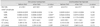
No significant differences were observed between RMTs obtained before and after daily rTMS in either the stimulated (p=0.379) or unstimulated (p=0.180) hemisphere (Table 1).
Recruitment curve
MEP amplitudes were smaller in the stimulated hemisphere after daily rTMS at intensities of 120% and 140% RMT (p=0.003 and <0.0001), but not in the unstimulated hemisphere (p=0.633 and 0.317). No differences were observed in MEP amplitudes at 100% RMT in both stimulated (p=0.146) and unstimulated (p=0.750) hemispheres.
Cortical silent period
Mean CSPs were prolonged in the stimulated hemisphere after daily 1-Hz rTMS (p=0.033). Similarly, the CSP tended to prolong in the unstimulated hemisphere (p=0.070).
SPM analysis of PET glucose metabolism
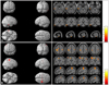
FDG-PET scans revealed a focal reduction in glucose metabolism in the stimulated area after 1-Hz rTMS for 5 days (Fig. 2A). However, despite the decreased TMS motor cortical excitability and increased cortico-cortical inhibition, glucose metabolism was not reduced in the primary motor cortex. On the other hand, glucose metabolism was markedly increased in the bilateral precentral, ipsilateral superior and middle frontal, prefrontal and cingulate gyri (uncorrected p<0.0001), which are remote from the stimulated area (Fig. 2B). Glucose metabolism was increased in the bilateral (more prominently ipsilateral) dorsolateral and ipsilateral medial frontal cortices to the stimulated hemisphere, indicating that these cortical areas might be related to the enhancement of cortico-cortical inhibition.
Discussion
The main findings of this study were (i) decreased MEP amplitudes at suprathreshold stimulation and prolongation of CSPs after daily 1-Hz rTMS, indicating a reduction of cortical excitability and increased ICI, and (ii) a focal reduction in glucose metabolism in the stimulated temporal cortex and increases in the bilateral precentral, ipsilateral superior and middle frontal, prefrontal and cingulate gyri. These findings suggest the 1-Hz rTMS affects not only the stimulated cortex but also remote cortical areas via modulation of cortical network properties.
Various effects of low-frequency rTMS of the frontal or prefrontal cortex are reported in the literature. One-Hz rTMS of the frontal motor or prefrontal cortex increased the RMT and decreased MEPs in normal healthy volunteers.6,7 However, few studies have investigated the effects of low-frequency rTMS of the temporal cortex. The choice of the temporal cortex as a stimulation target in this study was motivated by several factors: 1) few studies have stimulated the temporal cortex, with most instead using frontal or prefrontal stimulation, 2) the functional and structural anatomy of this area is well defined in humans, and 3) such studies are likely to have significant clinical implications given that the temporal cortex can be a target of treatments for tinnitus and temporal lobe epilepsy.4,6,8 Testing the feasibility and safety of repeated rTMS sessions was another important aspect of the present study, and these sessions were well tolerated, with only mild transient symptoms such as headache, tinnitus, and local irritation at the stimulated region occurring in three subjects. One recent study provided updated application guidelines for TMS use in clinical practice and research based on a review of safety concerns and side effects reported in the literature.7 One-Hz subthreshold rTMS was considered to represent acceptable and safe stimulation in both normal subjects and patients.
In the present study, the acute effects of daily 1-Hz rTMS were reduced MEP amplitudes at suprathreshold stimulation and prolongation of CSP in stimulated hemispheres. The variation of MEP amplitude with stimulation intensity (corresponding to RC) is enhanced by D-amphetamine and is influenced by GABAergic and monoaminergic neurotransmission and by the properties of Na+ and Ca2+ channels.24 We found that daily rTMS induced prolongation of CSPs in the stimulated hemisphere but did not alter ICI at an ISI of 2 ms, with both of these observations considered to reflect ICI. However, a different inhibition mechanism might mediate changes in CPS and ICI at a short ISI. In fact, the inhibition involved in the CSP and long-term ICI at ISIs from 50 to 200 ms is known to be related to GABAB activity,25 while short ICI (SICI) at ISIs of less than 5 ms are related to GABAA.26 The SICI and CSP clearly reflect different aspects of cortical inhibition, and 1-Hz rTMS of the temporal cortex seems to enhance inhibitory effects, espe-cially GABAB-related mechanism. A previous study that applied 1-Hz rTMS to motor cortices of healthy normal volunteers found a decrease in cortical excitability (increased RMT and decreased MEP amplitudes) at the stimulated motor cortex.6 It is well known that membrane potentials associated with changes in Na+ and Ca2+ channels prominently influence RMT,27 suggesting that 1-Hz rTMS decreased the membrane potentials of the stimulated neurons. In the present study, 1-Hz rTMS of the temporal cortex did not induce any changes in RMT, ICI, or ICF. This means that 1-Hz rTMS enhances GABAB-mediated inhibitory interaction rather than altering the cortical excitability of pyramidal neurons or the activity of excitatory interneurons. This type of stimulation may also influence neuronal activity in other cortical areas by modulating inhibitory interneurons. The relative proximity of the temporal cortex to the frontal cortex could have resulted in rTMS stimulating the primary motor cortex directly. However, changes in the CSP without MEP or RMT changes in the motor cortex of the unstimulated contralateral hemisphere indicate that focal stimulation of the temporal cortex can increase cortico-cortical inhibition while not influencing local neuronal membrane potentials. These findings could be considered to be strong evidence of cortico-cortical interactions induced by rTMS. We previously observed that the instantaneous EEG amplitudes in the alpha and beta frequency bands were increased after daily 1-Hz rTMS of the temporal cortex,9 but we did not observe any changes in TMS indices before daily rTMS in the current study. Daily accumulative effects were not observed either in the TMS indices.
Functional imaging has been used to investigate changes induced by focal rTMS in stimulated, adjacent, and remote cortical areas. One study showed that 2-Hz suprathreshold rTMS of the primary hand motor cortex increased glucose metabolism in the primary motor and premotor cortices.10 Another study using 15O-PET found that rTMS of the mid-dorsolateral frontal cortex modulated the cerebral blood flow (CBF) at the stimulated regions as well as at several cortical regions distal to anterior and posterior cingulate cortices.28 Other studies have found that 1-Hz rTMS of the primary hand motor area increased the CBF in the ipsilateral primary auditory, contralateral cerebellum, bilateral putamen, and red nucleus in an intensity-dependent manner, whereas there was an inverse correlation between the CBF in the stimulated and contralateral prefrontal cortices, ipsilateral medial temporal, both parahippocampal, and posterior middle temporal gyri during stimulation of the prefrontal cortex.11 Those authors suggested that the effects of 1-Hz rTMS on prefrontal cortex appear opposite to those of rTMS on the primary motor cortex, suggesting the presence of modulatory neural activity of the prefrontal cortex and other pathways downstream of that area. They also reported regional selectivity of the pathways stimulated, indicating functional connectivity between prefrontal and other cortical areas. However, none of these previous studies examined changes in cortical activity after rTMS of the temporal cortex. The presence of regional and remote effects of 1-Hz rTMS of the temporal cortex are supported by the PET changes in glucose metabolism observed in the present study. Our SPM PET results indicated that 1-Hz rTMS induced focal decreases in glucose metabolism, suggesting direct inhibitory effects on the underlying cortical neurons. If reduced glucose metabolism in the temporal cortex can be considered to result from decreased cortical neuronal activity in the stimulated area, what causes increased glucose metabolism in other cortical (i.e., bilateral precentral, ipsilateral frontal, prefrontal, and cingulate) areas that are remote from the stimulated cortex? These alterations could be considered to reflect enhanced cortico-cortical inhibition if the inhibitory activity was mediated by an active process as a result of functional activation in neuronal circuits involving cortico-cortical inhibitory interaction. Although the mechanisms underlying these remote effects cannot be interpreted in the setting of current study, anatomical and physiological mechanisms could be involved. The prefrontal and cingulate cortices are known as association cortical areas integrating cortical functions of other areas, such as decision making and/or inhibitory control on motor pathways.29,30
The temporal relationships of the rTMS effects have been assessed in the primate brain14 by performing FDG-PET serially before, during, and up to 16 days after 5-Hz rTMS of the precentral gyrus. Glucose metabolism decreased in the motor and premotor cortices and increased in the limbic-associated areas such as the cingulate and orbitofrontal cortices, and those changes continued for at least 8 days, suggesting relatively long-lasting effects of rTMS in the functionally connected cortical areas. A recent review article reported TMS-induced aftereffects in EEG and evoked potentials when various rTMS protocols and single sessions were applied to either healthy volunteers or patients.5 The mean aftereffect was a change of 30-35% from baseline or sham with a duration of less than 70 min (mean of 35 min), mainly in the alpha band and similarly for low-frequency (≤1 Hz) and high-frequency (≥5 Hz) rTMS, with only the effect direction (suppression vs. facilitation) varying with the number of pulses, pulse trains, and intensities used. Those authors were not aware of any study of differences in aftereffect following multiple rTMS sessions over consecutive days, but mentioned the importance of such studies especially for clinical applications.3,5 One study found that the effects of rTMS sessions on corticospinal excitability were stronger when they were applied 24 hours after the initial session, suggesting the presence of a neurophysiologic residual effect, although this was not strong enough to have clear behavioral or clinical implications.31 In the current study, the study design prevented further quantification of the precise durations of these changes, but future studies could attempt to determine the detailed time profiles of the effects of repeated stimulation for longer periods.
In summary, the present study has demonstrated that 1-Hz rTMS administered to the nondominant temporal cortex can reduce cortical excitability in the ipsilateral motor cortex and increase intracortical inhibitory interactions bilaterally, more prominently in the stimulated hemisphere. One-Hz rTMS also affects neural activities in motor and related cortical regions not only in the ipsilateral hemisphere but also in the contralateral hemisphere, possibly via modulations of intracortical interactions, as revealed by CSP changes and PET glucose metabolism analyses. Our results suggest that 1-Hz rTMS can acutely influence regional cortical activity, not only in the stimulated cortex but also in the adjacent and distant cortical areas, which modulates neuronal activity by affecting the long-range cortical network.
 XML Download