

 PDF
PDF ePub
ePub Citation
 Print
Print


Introduction
Transient ischemic attack (TIA) is defined as a reversible neurologic deficit that persists for less than 24 hours.1 It was originally assumed that this pathophysiology of TIA was associated with complete resolution of brain ischemia and that it would leave no permanent brain injury.2,3 However, due to advances in neuroimaging, it has been found that abnormalities are observed on diffusion-weighted imaging (DWI) for a substantial proportion of TIA patients.4-7 It has also been reported that the presence of DWI lesions is predictive of outcome in patients with TIA.8
Many TIA patients, irrespective of DWI lesions, have juxtacortical spots on fluid-attenuated inversion recovery (FLAIR) images. In general, small scattered lesions on DWI are considered to be small embolisms due to potential embolic sources such as carotid stenosis, intracranial atherosclerosis, or cardiac diseases. However, the clinical implications and etiologies of this phenomenon on FLAIR images have not been extensively researched in patients with TIA.
The foramen ovale remains open [patent foramen ovale (PFO)] in about one-quarter of the general population.9 An association between PFO and acute embolic lesions has been reported in patients with cryptogenic stroke or paradoxical embolism,10,11 and consequently there is an association between the presence of PFO and cryptogenic stroke.9 The etiology of TIA without arterial steno-occlusion or potential cardioembolic sources remains unclear. We hypothesized that TIA without clear etiology is partly associated with right-to-left shunt (RLS). Therefore, the aim of the present study was to determine whether there is an association between the presence of RLS and the occurrence of juxtacortical spots on FLAIR images in patients with TIA without clear etiology, and to compare the imaging findings of patients with and without RLS.
Methods
Subjects
This was a retrospective study of TIA patients who consecutively visited our tertiary stroke center via the outpatient department or the emergency department within 72 hours of TIA onset between October 2008 and October 2011. TIA was diagnosed according to the definitions given in the Classification of Cerebrovascular Disease III, National Institute of Neurologic Diseases and Stroke.1 We excluded the following patients from the study: 1) those with isolated vertigo without evidence of the central type, 2) those whose imaging or detailed evaluations were incomplete, and 3) those with other etiologies such as Moyamoya disease, autoimmune diseases, or hemiplegic migraine. This study was approved by the Institutional Review Board of our hospital. Written informed consent was neither obtained nor necessary because of the retrospective design of this study.
Clinical data and assessment of TIA symptoms
The patients were consistently evaluated according to our st-roke protocol, and the following clinical data were obtained for all patients: age, gender, and risk factors for stroke inclu-ding hypertension, diabetes mellitus, dyslipidemia, smoking, history of cardiac disease including atrial fibrillation, myocardial infarction, angina, valvular heart disease, and history of previous TIA. TIA symptoms were classified into the following: hemiparesis, monoparesis, sensory disturbance, facial palsy, dysarthria, ataxia, and cortical symptoms. Using the ABCD2 score criteria, we scored the following variables: age ≥60 years, 1 point; initial hypertension ≥140 mm Hg (systolic) and/or ≥90 mm Hg (diastolic), 1 point; weakness, 2 points; speech disturbance, 1 point; duration of symptoms >1 hour, 2 points; duration of symptoms from 10 minutes to 1 hour, 1 point; and diabetes mellitus, 1 point.12
Magnetic resonance imaging protocol and assessment
According to our stroke imaging protocol, patients underwent emergency magnetic resonance imaging (MRI) after admission and a follow-up DWI scan on day 3 or 4 if they had undergone DWI within 24 hours after symptom onset. The MRI protocol included axial DWI, gradient-echo imaging, FLAIR, and time-of-flight extracranial and intracranial angiography. Conventional MRI was performed with a 1.5-T system (Sigma, GE Medical Systems, Milwaukee, WI, USA) with echo-planar capabilities.
MRI findings were analyzed by two experienced stroke neurologists (J. T. K. and D. E. K.) who were blinded to the clinical data. Disagreements were resolved by consensus. Spots on FLAIR images were categorized as those in the subcortical area or juxtacortical area. Only juxtacortical spots were analyzed because deep white-matter hyperintensities (DWMHs) and periventricular white-matter hyperintensities (PVWMHs) could not be distinguished reliably. Juxtacortical spots were defined as small and round hyperintensities in the juxtacortical areas on FLAIR images, irrespective of recent DWI lesions and/or previously defined infarct lesions (Fig. 1). The presence and number of juxtacortical spots were assessed. In addition, we rated DWMHs as punctuate, early confluent, or large confluent lesions, and PVWMHs as caps and pencil-thin linings, smooth halo, or irregular PVWMHs extending into the deep white-matter according to Fazeka's Scale.13 If it was difficult to distinguish the spots of FLAIR images as lesions of extensive DWMHs (or PVWMHs) or juxtacortical spots, they were designated as DWMHs (or PVWMHs) rather than juxtacortical spots.
Arterial disease and cardiac workup
Magnetic resonance angiograms were analyzed and considered as stenosis if arterial stenosis had resulted in a 50% or greater narrowing of the lumen. Symptomatic arterial stenosis was considered as stenosis corresponding or contributing to the ischemic symptoms. All patients underwent 12-lead electrocardiography and 24-hour Holter monitoring. Transthoracic echocardiography or cardiac computed tomography was performed in patients without symptomatic arterial stenosis or those suspected to have cardiac diseases. We defined the cause of TIA and potential embolic sources according to the results of the diagnostic workup. TIAs with attributed causes of ischemia or potential embolic sources were designated as having a clear etiology. Patients without clear etiology or potential embolic sources despite routine diagnostic workup before a transcranial Doppler (TCD) with an agitated saline test or transesophageal echocardiography (TEE) were designated as having cryptogenic TIA.
The presence of RLS was established using TCD with an agitated saline test while the patient was at rest and while a Valsalva maneuver was being performed.14 This technique is carried out by injecting a mixture of saline with air into the antecubital vein and concomitantly recording the blood flow velocity in the right (or left) middle cerebral artery or basilar artery. If there is an RLS, the contrast medium causes typical high-intensity transient signals during contrast TCD monitoring of the middle cerebral artery. The degree of shunting was defined as follows: 1) no microbubble; 2) grade 1, 1-10 microbubbles; 3) grade 2, >10 microbubbles and no curtain; and 4) grade 3, curtain or shower of microbubbles.14 In patients with PFO or suspected PFO, TEE with an agitated saline test was also performed. If there was a discrepancy between TCD and TEE, the presence of PFO was decided by a consensus meeting of a neurologist and a cardiologist, who was not involved in this study.
Statistical analysis
Cryptogenic TIAs were further divided into RLS-positive and RLS-negative subgroups, and the differences in imaging findings were compared between these subgroups. Data are presented as mean±SD values or as the frequencies of categorical variables. The chi-square test or Fisher's exact test was used for categorical variables, and the Mann-Whitney U test and the Kruskal-Wallis test were used for continuous variables in univariate analyses. Multiple logistic regression analysis was used to evaluate the independent factors associated with juxtacortical spots, and included in a logistic regression model in which the entry was set at a univariate association of p<0.2. The level of statistical significance was set at p<0.05. SPSS for Windows (version 17.0, SPSS, Chicago, IL, USA) was used for all statistical analyses.
Results
Baseline characteristics
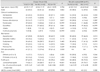
Of the 256 patients with TIA who were screened, 29 were excluded: 4 because of intravenous thrombolysis, and 25 because of incomplete diagnostic workups (5 imaging, 10 cardiac, and 10 RLS workups). Among the remaining 227 patients, 132 (66 men, 66 women; age, 60.35±11.37 years) were designated as having cryptogenic TIA, and 41 of those patients (31.1%) had DWI abnormalities (Table 1). Seventy (53.0%) of the cryptogenic TIA patients had RLS, which was diagnosed in 39 patients by both TCD and TEE, in 29 by TCD alone, and in 2 by a consensus meeting because of a discrepancy between the 2 diagnostic methods. Patients with RLS presented more frequently with hemiparesis than those without, but ABCD2 scores did not differ significantly between patients with and without RLS (p=0.084). Ninety-one patients without lesions on DWI presented similar results.
Imaging characteristics of the cryptogenic TIA patients
DWI abnormalities and juxtacortical spots on FLAIR images were detected more frequently in patients with RLS (41.4% and 71.4%, respectively) than in those without (19.4% and 38.7%, respectively). The number of juxtacortical spots was also higher among patients with RLS than in those without. The severity of white-matter hyperintensities (as assessed using Fazeka's score) did not differ significantly between patients with and without RLS. The results were similar for the 91 patients without lesions on DWI.
Spots on FLAIR images were more frequently observed in patients with DWI abnormalities and RLS than in those without. Juxtacortical spots were also associated more with older age than with no spots (Supplementary Table 1). The independent factors associated with juxtacortical spots on FLAIR images were RLS [odds ratio (OR)=3.802, 95% confidence interval (95% CI)=1.74-8.2; p=0.001] and age (OR=1.058, 95% CI=1.01-1.10; p=0.004) by multivariate analysis. The variables tested in a multivariate logistic regression model were age, presence of DWI lesions, ABCD2 score, and presence of RLS (p<0.2 by univariate analysis in Supplementary Table 1). RLS was independently associated with juxtacortical spots on FLAIR images even after excluding patients with DWI lesions (n=91; OR=4.297, 95% CI=1.71-10.82; p=0.002; Table 2).
The frequency and number of juxtacortical spots on FLAIR images were significantly higher in patients with severe RLS (grades 2 or 3) than in those without RLS (p<0.001, Table 3).
Discussion
In this study we found an association between the presence of RLS and juxtacortical spots on FLAIR images in TIA without clear etiology. There was a strong correlation between the degree of shunting and the number of juxtacortical spots on FLAIR images. To the best of our knowledge, few studies have assessed the association between TIA without clear etiology and RLS and compared the differences in imaging findings between TIA patients with and without RLS.
The diagnosis of PFO was obtained for about 60% of the patients using TEE. Since RLS might also be caused by pulmonary shunting in a small proportion of patients, we considered the presence of RLS instead of PFO in this study. However, it has recently been demonstrated that TCD with an agitated saline test may be a good tool for the detection of PFO; it has a similarly good sensitivity and specificity to TEE.15,16 Our study also showed that TCD with an agitated saline test and TEE were similarly effective for detecting RLS.
Since it is often difficult in practice to distinguish juxtacortical spots on FLAIR images from severe DWMHs, we accepted only clear, isolated juxtacortical spots that were distinct from any DWMHs, and divided them into juxtacortical and cortical-subcortical lesions. We presumed that juxtacortical spots on FLAIR images represented a silent and/or subclinical embolism. However, a new definition of silent spots on FLAIR images is needed and their clinical implications tested in the future. In previous studies, a silent embolism has been described as a DWI lesion without clinical symptoms.17 Several studies suggest that acute silent embolisms are associated with RLS (or PFO).18 Our results support the hypothesis that silent DWI lesions are associated with juxtacortical spots. However, further study is needed to confirm this hypothesis.
One possible explanation for the finding that an embolism arising via RLS is usually silent or transient is that the embolism is small and washed out under normal physical conditions. Venous emboli arising via RLS might be susceptible to intrinsic fibrinolysis;19 thus, in patients without potential embolic sources, the presence of multiple juxtacortical spots on FLAIR images suggests that RLS is the source of a subclinical embolism, but that the symptoms could be associated more with clinical parameters such as age, heart function, or coagulation function. An association between the presence of PFO and cryptogenic stroke has been reported.9 However, the association between RLS and DWI abnormalities in TIA has not yet been demonstrated in previous studies.7 The findings of the present study are different from ours in that they included general patients with TIA. Since RLS was considered a low-to-medium risk for embolism, we considered that it could also be a possible risk factor for TIA only after excluding patients with clear etiologies such as potential cardioembolic sources or arterial steno-occlusion. TIA patients with clear etiologies could have a higher frequency of juxtacortical spots on FLAIR images and DWI lesions compared to those without clear etiologies. Since we included only patients without a clear etiology, RLS could be considered an important factor for juxtacortical spots on FLAIR images.
Juxtacortical spots on FLAIR images warrant further investigation. Previous studies have demonstrated that PVWMHs and DWMHs are significantly related to recurrent stroke, poor outcome, hemorrhagic transformation, and cognitive decline. We are currently working to establish whether the risk for recurrent stroke is higher in our patients than in others. Based on the results of previous studies, it is conceivable that the risk of recurrent stroke may be higher in patients with juxtacortical spots than in those without such spots. Although it has been determined that PFO is not correlated with recurrent stroke, the findings for patients with both PFO and juxtacortical spots may be different. Thus, the results of our study provide the basis for future investigations, and additional prospective studies are warranted.
Study limitations
This study was subject to several limitations due to its retrospective design and small sample. In addition, we performed an RLS study only in cases for which the exact cause could not be determined after routine diagnostic workups. Thus, we could not find an association between all patients with TIA and RLS. In a previous study, only RLS with atrial septal aneurysm was found to be closely associated with cryptogenic stroke.9 However, atrial septal aneurysm was detected infrequently in the present study (n=3, 4.2%). Furthermore, while the definition of juxtacortical spots has been applied to migraine patients in previous studies,20 this is the first time it has been applied to TIA. In contrast to a previous migraine study, the present study involved patients of older age, for which white-matter hyperintensities are common and could have led to a falsely high discovery of spots on FLAIR images. While spots that were considered to be due to severe white-matter hyperintensities were excluded as juxtacortical spots, these criteria may have influenced the results. Furthermore, silent lesions of multiple sclerosis are known to manifest as juxtacortical spots; however, it is unlikely in the present population, since the incidence of multiple sclerosis is very low in Korea.
Conclusion
There may be a significant association between the presence of RLS and juxtacortical spots on FLAIR images in TIA patients without clear etiology. The results presented here suggest that RLS is one possible source of lesions on FLAIR images in cryptogenic TIA. Further study is needed to confirm these results, to improve our understanding of the association between TIA and RLS, and to establish the clinical implications of juxtacortical spots on FLAIR images.


 XML Download