

 PDF
PDF ePub
ePub Citation
 Print
Print


Introduction
It is being increasingly recognized that the prevalence of vascular cognitive impairment (VCI) is higher than originally thought. Since VCI is considered to develop under the influence of various cardiovascular risk factors, it is emerging as a potentially treatable and preventable form of dementing disorders.1 However, currently available therapeutic options for treatment of dementia, such as cholinesterase inhibitors, were originally devised for Alzheimer's disease (AD), and their application in vascular dementia (VaD) has produced limited and uncertain clinical benefit.2
It is widely recognized that at the incipient stage, AD patients generally complain about memory problems, whereas VaD patients usually present with cognitive deficits involving nonmemory domains.3 However, a substantial proportion of stroke survivors suffer from poststroke memory dysfunction, and several factors, including medial temporal atrophy (MTA) or white matter hyperintensities (WMHs), are associated with poststroke memory dysfunction.4 Furthermore, there is increasing evidence that coexisting cerebrovascular disease precipitates and unmasks underlying preclinical AD.5 Stroke and AD are highly prevalent and the two conditions share common vascular risk factors, and so it can be hypothesized that patients with poststroke memory dysfunction may have had subclinical AD before their stroke, and their memory deficits may be the divulgence of underlying AD triggered by stroke. This issue has not been studied in populations with cognitive impairment-no dementia (CIND), which is clearly the foremost target of therapeutic interventions for preventing dementia.6 We sought to test this hypothesis by measuring the severity of MTA, which is an acknowledged neuroimaging index of AD.7 For this purpose, we compared patients with poststroke amnestic vascular cognitive impairment-no dementia (aVCIND) and patients with nonstroke amnestic mild cognitive impairment (aMCI) to patients with poststroke nonamnestic vascular cognitive impairment-no dementia (naVCIND).
Methods
Subject recruitment
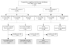
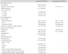
This was a retrospective analysis based on a prospective cognitive test database. The patient selection process is summarized in Fig. 1. A battery of standard neuropsychological tests was administered to 1201 patients at Seoul National University Bundang Hospital between May 2007 and March 2009. Of these 1201 patients, 396 had a documented history of clinical stroke with which neuroimaging findings were compatible; the remaining 805 patients were designated as nonstroke subjects. Stroke was defined using the World Health Organization definition of "rapidly developed clinical signs of focal or global disturbance of cerebral function, lasting more than 24 hours or leading to death, with no apparent cause other than a vascular origin".8 CIND was defined as having cognitive impairment but not meeting the Diagnostic and Statistical Manual of Mental Disorders, Fourth Edition, Text Revision (DSM-IV-TR) criteria for dementia.6,9 We also applied an Instrumental Activity of Daily Living score of <0.43 as indicating "no dementia".10
For poststroke patients, a standardized neuropsychological battery was administered at least 90 days after stroke onset. The interval from stroke onset to neuropsychological evaluation was 473±311 days (mean±SD). We selected poststroke VCIND patients (n=156) who had an impairment in one or more cognitive domains but did not meet the DSM-IV-TR criteria for dementia. Cognitive impairment in language, visuospatial function, or memory domains was defined as a score in the each of the domain-specific tests of less than the 7th percentile (mean-1.5 SD). Cognitive impairment in the frontal domain was determined by a score of less than the 7th percentile in two or more of the five frontal-domain-specific tests (details of the neuropsychological tests used are given below). Post-stroke VCIND was further classified into poststroke amnestic VCIND (aVCIND; n=71) and poststroke nonamnestic VCIND (naVCIND; n=85), according to the presence of memory impairment. Among 805 nonstroke patients, 331 were identified as having CIND; 128 patients who had cognitive impairment in the memory domain were further categorized into the aMCI group. Patients without coronal magnetic resonance imaging (MRI) results were excluded (23 from the aVCIND group, 35 from the naVCIND group, and 63 from the aMCI group). These criteria resulted in 163 patients (48 aVCIND, 50 naVCIND, and 65 aMCI) being included in this study. All of the included patients were free of any other organic medical or neurological conditions that may adversely affect cognitive functions. The study protocols were approved by the local institutional review board.
Cognitive assessment
A battery of standard neuropsychological tests was adapted from the 60-minute neuropsychology protocol of the Vascular Cognitive Impairment Harmonization Standards (VCIHS).11 The Korean-VCIHS Neuropsychologic Battery was standardized for the Korean population, and its validity and feasibility were examined and confirmed in a multicenter epidemiological study involving 15 hospitals with nationwide coverage.12 The Korean-VCIHS Neuropsychologic Battery comprises the following cognitive tests of various cognitive domains: frontal executive/activation (animal naming test, phonemic fluency test, Digit Symbol Coding, Trail Making Test), language/lexical retrieval (Korean-Boston Naming Test - short form), visuospatial (Rey-Osterrieth Complex Figure Test: Copy), memory (Seoul Verbal Learning Test), depressive mood (Geriatric Depression Scale), and others (Informant Questionnaire for Cognitive Decline in the Elderly, Korean Mini Mental State Examination, and Instrumental Activity of Daily Living). Trained clinical psychometricians who were blinded to the clinical and radiological profile of each patient administered the battery. The score on each cognitive test was transformed into a standardized Z-score [Z-score=(individual score - population mean score)/(population SD)].
Vascular risk factors and MRI evaluation of the patients
The baseline demographic and clinical characteristics of patients included in the study were collected, including age, gender, years of education, handedness, and vascular risk factors, such as presence of hypertension (previous use of antihypertensive medication, systolic blood pressure >140 mm Hg, or diastolic blood pressure >90 mm Hg), diabetes mellitus (previous use of glucose-lowering medication, fasting blood glucose >7.0 mmol/L, or 2-hours-postprandial blood glucose >11.1 mmol/L), dyslipidemia (previous use of lipid-lowering medication, total cholesterol >6.0 mmol/L, or low-density lipoprotein cholesterol >4.14 mmol/L), and smoking (current smoker or stopped smoking within the past 5 years). For post-stroke patients, the severity of stroke was assessed using the National Institutes of Health Stroke Scale score, which was measured at hospital admission for the most-recent stroke, and the modified Rankin Scale score, measured at discharge from that admission.
All participants underwent brain MRI, and the mean interval between MRI and test for the neuropsychological battery was 153 days. The MRI studies were performed using a 1.5-tesla superconducting magnet (Intera, Philips Healthcare, Eindhoven, The Netherlands). The standardized MRI protocols consisted of an axial T2-weighted spin echo, coronal T2-weighted spin echo, fluid-attenuated inversion recovery image, gradient-echo image, and axial T1-weighted image. MTA was rated using a 5-point rating scale, as described previously.13 Both sides were rated simultaneously, and in the case of noticeable asymmetry, the score of the more affected side was chosen as being representative.14 WMHs were rated visually using a 10-point rating scale, based on the fluid-attenuated inversion recovery image.15 Information regarding the subtype, location, and laterality of the stroke and the involvement of the medial temporal lobe was obtained from the prospective stroke registry and by reviewing the electronic medical records and MRI findings. Two reviewers (B.J. Kim and M.-Y. Oh) independently and blindly rated the MTA and WMHs (Spearman's correlation coefficient: 0.79 for MTA and 0.73 for WMHs), and any disagreement was settled by consensus.
Statistical analysis
Differences in baseline characteristic variables among the poststroke aVCIND, poststroke naVCIND, and aMCI groups were examined by using Pearson's χ2 test, the Kruskal-Wallis test with pairwise Mann-Whitney U test, or one-way analysis of variance with Bonferroni's post-hoc adjustment for multiple comparisons, as appropriate. Z-scores were described as mean±SE values. Bivariate analyses and multivariable ordinal logistic regression analyses performed by taking the MTA score as a dependent variable were used to assess whether covariates were associated with an increasing severity of MTA. To build ordinal logistic regression models, three patients with the most-severe MTA (MTA score=4) were combined with the group of patients with an MTA score of 3. The distribution of patients by MTA score used in the model was as follows: 60 (36.8%), 52 (31.9%), 29 (18.0%), and 22 (13.5%) patients with MTA scores of 0, 1, 2, and 3 or 4, respectively. The proportional odds assumption for each ordinal logistic regression model was examined and found to be satisfactory. Variables with a bivariate p<0.10 for their association with MTA were selected for adjustment in multivariate models, including age, WMHs, hypertension, and diabetes. Statistical significance was defined as a probability value of p≤0.05. All statistical analyses were performed using SPSS 15.0 (SPSS, Chicago, IL, USA).
Results
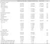
The distributions of gender and MTA differed significantly among the three CIND groups; however, age, dominant hand, education, WMHs, and cardiovascular risk factors did not differ (Table 1). Stroke-related features, the location and laterality of stroke lesions, the stroke subtype, and the severity of the stroke did not differ significantly between poststroke aVCIND and naVCIND patients. The MTA score varied significantly among the three types of CIND (p<0.01, Kruskal-Wallis test). Pairwise comparisons revealed that the MTA score was significantly lower in the poststroke naVCIND group (median, interquartile range; 1, 0-2) than in the poststroke aVCIND group (1, 0-1; p=0.022) and the aMCI group (1, 0-2; p<0.01; the significance level was set to p≤0.017 after adjusting for pairwise comparison). However, the MTA score did not differ between the poststroke aVCIND and aMCI groups (p=0.10).
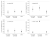
Analysis of variance showed that with the exception of the memory score, the domain-specific test scores did not differ among the three CIND groups (Table 2) (Fig. 2). The memory domain was used to differentiate poststroke aVCIND from poststroke naVCIND. Global Frontal Function score, obtained by averaging the Z-scores of the five frontal domain-specific tests, did not differ between the groups (p=0.94).
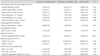
Ordinal logistic regression analyses showed that there was a significant association between the severity of MTA and the type of CIND (Table 3). In multivariable analysis, the odds ratio for poststroke aVCIND was 2.69 (95% confidence interval=1.21-5.99) and the odds ratio for aMCI was 5.20 (95% confidence interval=2.41-11.23) relative to poststroke naVCIND. When compared with aMCI, poststroke aVCIND was not significantly associated with the severity of MTA (p=0.11). In addition, age and diabetes mellitus were independently associated with increasing severity of MTA (Table 3).
Discussion
The patients in this study were divided into two groups according to the presence of memory dysfunction so as to make it possible to evaluate the association between MTA and memory dysfunction in patients with poststroke CIND. The effect of stroke was evaluated by comparing nonstroke aMCI patients with poststroke CIND patients. With the exception of memory, patients in the three CIND groups defined in our study had similar cognition and functional ability profiles. We found that the odds of having more-severe MTA were increased for poststroke aVCIND and nonstroke aMCI relative to poststroke naVCIND. Older age and diabetes mellitus were also found to be independently associated with MTA.
The concept of poststroke memory dysfunction seems counterintuitive in that stroke does not usually affect the memory-processing structures.16 However, one-third to one-half of stroke survivors were reported to have memory dysfunction around 3 months after the stroke,4 and the risk of dementia doubles with a history of stroke.17 Both MTA and imaging markers of cerebrovascular disease are useful in predicting the development of clinical dementia.18 Experimental studies have also suggested that certain mechanisms are responsible for progressive memory deterioration. Decreased cerebral blood flow, which occurs with ischemic stroke, may modulate trafficking of β-amyloid between the blood and the brain, resulting in the attenuation of β-amyloid clearance from the brain.19,20 Moreover, small ischemic injuries that develop in the paraventricular area and deep structures may cause widespread disconnection of cholinergic innervations to the cortex,21,22 thereby inducing additive attrition of cognitive reserve.23
We deliberately recruited CIND patients from our large population of patients with neuropsychological assessments. Cholinesterase inhibitors and memantine, which are used to treat AD patients, seem to provide little benefit to VaD patients,2 and there has been no clinical trial regarding their efficacy in the treatment of poststroke dementia. According to DSM-IV-TR criteria, the presence of stroke per se in a demented individual excludes a diagnosis of AD. However, our results, as well as those from other recent studies, show that VaD patients usually have "mixed" pathology.24,25 In this context, our findings suggests that poststroke CIND can be regarded as subclinical AD if there is MTA or memory dysfunction, and that cholinesterase inhibitors or memantine can be beneficial for these individuals. In addition, the consideration of MTA when recruiting subjects for VCI trials or in secondary analysis of VCIND research would improve the homogeneity of study populations.
The association between MTA and poststroke dementia has been addressed by previous studies. However, the clinical implications of the results obtained in those studies were limited because they disregarded CIND or were case series of stroke at a specific location.26-29 In addition to the type of CIND, our study detected increases in the severity of MTA with the presence of diabetes, which is consistent with a previous report.30 WMHs and hypertension were not associated with severity of MTA in multivariable analyses, in spite of their significance in univariate analyses (Table 3). A lack of power due to the small sample is a possible explanation for this finding. Furthermore, previous studies also found that the association between WMHs and MTA is controversial.31,32 Contrary to the concept of early executive dysfunction in VaD, frontal function did not differ among the three CIND groups in the present study. However, the development of executive dysfunction has been reported to be as common in AD as in VaD.33
There are several reasons why our results should be interpreted with caution. First, this study was conducted at a single center, had a cross-sectional design, and a relatively small sample. Second, it should be noted that approximately 5% of MCI patients are reported to return to normal over time and that the natural course of progressive memory deterioration is thought to be dynamic.4,34 Third, single- and multidomain CINDs were not distinguished in our study due to the small sample. Fourth, the visual rating scale of MTA was used instead of a volumetric assessment. However, volumetry is cumbersome because it requires expensive software and a time-consuming rendering process, which limits its usefulness for research purposes. Moreover, it has been reported that visual and volumetric assessments of MTA are equally accurate.35,36 Fifth, prestrike neuropsychological assessments were not available for our subjects.
Our results do not indicate that poststroke memory dysfunction is essentially AD or that such patients should receive cholinesterase inhibitors - only a prospective cohort study with an adequate duration of follow-up can clarify the association between AD and poststroke memory dysfunction. Considering the increasing burden of stroke and dynamic interrelationships among vascular etiologies, degenerative changes in the brain, and host factors with respect to VCI,37,38 our findings support the notion that a stroke event may trigger subclinical AD-type neurodegeneration and result in poststroke memory dysfunction.
 XML Download