

 PDF
PDF ePub
ePub Citation
 Print
Print


Introduction
The term "EMG disease" is used to describe the unexpected finding of diffusely increased insertional activity on needle electromyography (EMG) in the absence of neuromuscular disease.1 Other names synonymous with this entity include "syndrome of diffuse abnormal insertional activity" and "syndrome of diffuse positive waves".2
EMG disease was first described in 1979 by Wiechers and Johnson,1 who reported on ten otherwise asymptomatic patients with short trains of provoked positive sharp waves (PSWs) in all muscles studied. Wright et al.3 reported on a family group with EMG disease with apparent autosomal dominant transmission and female predominance. Chow et al.2 reported on a patient with EMG disease that involved the bulbar, trapezius, frontalis, and orbicularis oculi muscles. It was recently suggested that this entity lies within the phenotypic spectrum of myotonia congenita (MC). Mutations in CLCN1, which encodes the human skeletal muscle chloride channel that contributes mainly to resting membrane stability, has been reported in two patients with EMG disease.4
EMG disease is rare, and prior published descriptions refer mainly to small numbers of patients. Therefore, the clinical characteristics of EMG disease have not yet been precisely elucidated. The aims of the current study were to determine the clinical characteristics of EMG disease and the relationship between EMG disease and CLCN1 mutations.
Methods
Subjects
All patients with EMG disease, which was diagnosed by needle EMG, were recruited between April 2008 and June 2011 from our neuromuscular laboratory clinics. Only EMG-disease patients with more than 1 year of follow-up were enrolled for this study. EMG disease was defined as diffusely increased insertional activity on needle EMG examination in the absence of any clinical or laboratory evidence of a known neuromuscular disease.1-4 In the current study, the electromyographic criteria of EMG disease was as follows: 1) sustained form of provoked PSWs with the time of insertional activity more than 100 ms after needle movement cessation for monopolar needle electrodes, bearing in mind that the electrical activity could continue for 48±18 ms (mean±standard deviation) following the cessation of electrode insertion into normal muscle tissues,5 and no insertional activity that extended for longer than 100 ms;6 2) presence at essentially every muscle tested including proximal and distal arm and leg, or paraspinals;1-4 3) absence of typical myotonic discharges that fire repetitively at 20-100 Hz and exhibit wax and wane variability in amplitude and frequency; and 4) motor-unit action potentials (MUAPs) with normal size, shape, number, and configuration. Informed consent to participate in this study was obtained from all enrolled patients, and the project was approved by the institutional review board.
Methods
The clinical characteristics of EMG disease were established by evaluating the clinical features of each enrolled patient by obtaining the following information: age, gender, time interval from symptom onset to diagnosis, site and aggravating factors of muscle stiffness, the presence of action and percussion myotonia, the presence of myalgia (muscle cramps), underlying diseases, and the results of a serologic examination including muscle enzymes (creatine kinase, myoglobin, alanine aminotransferase, aspartate aminotransferase, lactate dehydrogenase, and aldolase), electrolytes (sodium, potassium, chloride, and total and ionized calcium and magnesium), thyroid function, and tumor markers. Genetic testing for myotonic dystrophy type I was also performed.
Underlying neuropathy was ruled out by performing nerve conduction study (NCS) using standard techniques.7 The presence of EMG disease was investigated by conducting an EMG study of both the upper and lower extremities, as well as the cervical (C5-T1) and lumbar (L2-S1) paraspinal muscles. Six proximal limb muscles (deltoid, biceps brachii, triceps brachii, gluteus maximus, iliopsoas, and rectus femoris) and five distal limb muscles (first dorsal interossei, abductor pollicis brevis, extensor digitorum communis, tibialis anterior, and medial gastrocnemius) were examined. Follow-up electromyographic examinations were performed about 6 months after the initial needle EMG studies, which were performed using monopolar needle electrodes by a single expert electromyographer.
To determine any mutations of CLCN1 associated with MC, genomic deoxyribonucleic acid (DNA) was extracted from peripheral blood using a standard protocol, and all coding regions of CLCN1 (exons 1-23; exons numbered according to RefSeq NM_000083.2, GenBank) were amplified by the polymerase chain reaction (PCR). PCR assays were carried out using 1.25 U of AmpliTaq Gold DNA polymerase (Applied Biosystems, Foster City, CA, USA), 100 ng of genomic DNA, 2.0 mM MgCl2, and a set of primers at 10 µM (data not shown in detail). The amplification conditions for each exon of CLCN1 were as follows: initial denaturing cycle at 95℃ for 5 min, followed by 35 cycles of denaturation at 95℃ for 30 sec, annealing at 63℃ for 45 sec, and extension at 72℃ for 1 min, followed by a final extension step of 72℃ for 5 min. The PCR products were electrophoresed on a 1.2% agarose gel and the amplified genomic DNA fragments were extracted from the gel and purified using the manufacturer's recommended protocol (QIAquick gel extraction kit, Qiagen, Hilden, Germany). Direct sequencing of both strands was performed with the aid of BigDye terminator chemistry (PE-Applied Biosystems), and each electropherogram visually analyzed using ChromasPro 2.13 (http://www.technelysium.com.au/chromas.html). The effect of the documented amino acid substitution on CLCN1 was obtained by PolyPhen-2 (http://genetics.bwh.harvard.edu/pph2/).
Results
Clinical characteristics
Six Korean patients with EMG disease were enrolled in this study. The patients ranged in age between 20 and 72 years; four (67%) were men and two (33%) were women. The median onset age was 26 years (range, 10-58 years) and the median time interval from symptom onset to diagnosis was 12.5 years (range, 1-27 years). The time from diagnosis to the last follow-up was 22.3±6.7 months (range, 13-31 months).
Three patients [subject number (SN)-I, SN-IV, and SN-VI] with a history of generalized myalgia were referred to our hospital to be evaluated for the possibility of motor neuron disease, because their needle-EMG-provoked PSWs were diffusely documented at all muscles studied. Another patient (SN-II) with chronic muscle stiffness at both upper extremities, who had fully recovered from poliomyelitis of the left arm, underwent EMG for evaluation of postpoliomyelitis syndrome. The remaining two patients (SN-III and SN-V) simply had a history of chronic, generalized myalgia.
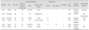
There was no specific clinical evidence of motor weakness, sensory loss, or muscular hypertrophy/atrophy among the six enrolled patients, and their muscle stretch reflexes were normal and bilaterally symmetric. The most consistent clinical feature was diffuse and chronic muscle stiffness of the upper and/or lower extremities, which was aggravated by exposure to cold. Immersing the patients' hands or feet in cold water aggravated the muscle stiffness without weakness. Furthermore, the muscle stiffness could not be relieved by muscle stretching, soft massage, or prescribed drugs, including carbamazepine, phenytoin, clonazepam, and mexiletine. Four patients (SN-I, SN-III, SN-IV, and SN-V) complained of slow relaxation of their hand muscles after voluntary contraction several times a year, but the symptoms never interfered with routine physical activity. Generalized myalgia was observed in four patients (SN-I, SN-III, SN-V, and SN-VI). However, muscle enzymes including creatine kinase (as a measure of sarcolemmal integrity) were within normal limits. Other laboratory test results for metabolic and electrolyte abnormalities were also unremarkable. In addition, searches for an underlying internal malignancy were negative. There was no common factor in their underlying diseases. The clinical characteristics of the subjects are listed in Table 1.
Electrophysiological characteristics
There was no evidence of peripheral neuropathy on routine NCS of one upper and both lower extremities in any of the patients. However, a needle EMG study in all patients revealed the continued presence of provoked PSWs with a time of insertional activity of more than 100 ms (duration, typically 200-1600 ms), which were found diffusely throughout the upper and lower extremities and in the paraspinal muscles (supplemental Data-Video file). The PSWs were identical to those seen in abnormal neurogenic, myogenic, and metabolic conditions. No fibrillation, fasciculation, or myotonic discharges were documented, although some of the patients had symptoms suggestive of action myotonia. MUAPs were of normal size, configuration, and number in all enrolled patients. In two patients (SN-II and SN-IV) with chronic muscle stiffness of the upper arms, increased insertional activities were identified in the asymptomatic muscles of both legs and the paraspinalis muscles. They were also recorded in the cranial muscles, such as the frontalis, masseter, and trapezius, in patient SN-I.
Genetic testing for CLCN1 mutation
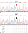
Direct sequence analysis of 23 exons of CLCN1 associated with MC revealed a mutation in only 1 of the 6 patients: patient SN-V carried a heterozygous substitution of T to C in exon 15 at position 1679 that caused an amino acid change at codon 560 (methionine to threonine, Met560Thr) (Fig. 1A), which was not found in 200 normal controls from South Korea or in the patient's asymptomatic mother or sister (Fig. 1B). This p.M560T mutation exchanged highly conserved amino acid residue in CLCN1 that has been evolutionarily conserved from zebrafish to humans (Fig. 1C). The pathogenicity of p.M560T mutation was also supported by PolyPhen-2, which predicted to be probably damaging with a score of 0.998 and not tolerated.
Discussion
EMG disease is characterized by diffusely increased insertional activity, which is seen as short trains of PSWs after the initial burst when needle movement stops. Insertional activity may be increased in various neuromuscular disorders in which there is irritability of the muscle fiber membrane.8 Although PSWs are not synonymous with the denervation process, their presence is usually associated with some type of pathology or blocked fibrillation potential.8 However, Wiechers and Johnson1 were the first to report cases with diffuse abnormal electromyographic insertional activity in the absence of any clinical or laboratory evidence of a known neuromuscular pathology. Johnson later coined the term EMG disease because the only abnormality is electrical.2
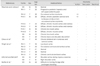
The clinical signs and symptoms of EMG disease in the previously cited articles are not well described, presumably because the related studies may have focused on the characteristic EMG findings. Furthermore, EMG disease may be quite rare, as seen in a previous study.3 It has not been observed any other cases in over 5000 EMG studies performed in their laboratory, with the exception of 1 family group.3 In the current study we found that patients with EMG disease shared the characteristic complaint of diffuse and chronic muscle stiffness or generalized myalgia that is exacerbated during cold exposure. The presence of cold sensitivity might be related to the type of CLCN1 mutation, although this has generally been considered a unique feature of paramyotonia congenita.9,10 Wiechers and Johnson1 reported that seven out of ten (70%) enrolled patients with EMG disease complained of chronic muscle aches and/or low-back aches. Chow et al.2 reported a case of EMG disease with bulbar muscle involvement who also showed restricted cervical range of motion. In addition, Nutter et al. reported on an EMG-disease patient who had difficulty moving his legs after prolonged standing.11 The clinical characteristics of subjects in these published descriptions are summarized in Table 2, and they are not considerably different from the symptoms and signs of our study subjects. Wright et al.3 found autosomal dominant transmission in an EMG study of a family group.
Some authors believe that EMG disease may be a mild form of or within the same spectrum as MC. It has been reported that two patients with EMG disease had known heterozygous substitutions (C2680T and C1649T) in CLCN1.4 However, we are not certain whether EMG disease is actually a form of MC for the following reasons: 1) only one CLCN1 mutation was identified in the current study; 2) none of our subjects had pathologic manifestations including muscular weakness or dystrophic features; 3) no electromyographic evidence of typical myotonic runs was found at rest or during volitional activity, even in the follow-up EMG study; and 4) no subjects in this study had a family history of MC.
It has recently been reported that absent, minimal, or waning-only myotonic discharges can be recognized on the initial EMG in patients with myotonic dystrophy type 2 (DM2),12 which is inherited by an expansion of the CCTG repeat in intron 1 of ZNF9/CNBP on chromosome 3.13 DM2 often presents with nonspecific symptoms such as muscle stiffness, myalgia, fatigue, or asymptomatic creatine kinase elevation.14,15 Three of the EMG-disease patients in the current study had a history of chronic muscle stiffness, clinical myotonia, or chronic myalgia, which can be suspicious of DM2. Therefore, the possibility of DM2 could not be excluded completely in patients without the genetic evidence of a CLCN1 mutation.
The results of the current study are subject to several limitations: 1) a relatively small study population was enrolled, 2) the follow-up duration of 1 year was rather short to search for hidden neuromuscular disease, 3) direct sequence analysis was limited to the coding regions and the exon-intron boundaries in CLCN1, and 4) additional genetic testing including of CCTG-repeat expansion in ZNF9 was not performed.
In conclusion, the clinical features of patients with EMG disease might be quite similar to those of MC patients. However, mutations in CLCN1 might be identified in only a few subjects. Further clinical and genetic studies are needed to verify the etiology and clinical features of EMG disease.
 XML Download