

 PDF
PDF ePub
ePub Citation
 Print
Print


Introduction
Sleep deprivation (SD) is known to cause profound impairments in executive function and vigilant attention.1,2 SD is reported to be associated with increases in blood pressure and stress hormone levels, and a decrease in parasympathetic tone.3,4 Stress hormones are regarded as factors that can interfere with cognitive function, including memory5; it has been shown that the increased levels of adrenal steroids induced by psychological stress blocks hippocampal long-term potentiation.6 The present study was designed to investigate whether 24 h of SD negatively affects the attention and working memory and increases the serum concentrations of stress hormones, glucose, and inflammatory markers.
Methods
Subjects and study protocol
Six normal healthy men (mean age 25.3 years, range 23-27 years; mean duration of education 13.5 years, range 14-18 years) participated in the study. All subjects were good sleepers and non habitual nappers with no excessive daytime sleepiness, and they had low anxiety levels. Sleep quality and quantity were assessed by questionnaires and actigraphy recordings. The subjects performed a cognitive test [the Continuous Performance Test (CPT)] in the morning after a normal night of sleep and then again after 24 h of SD. Serum concentrations of stress hormones (cortisol, epinephrine, and norepinephrine), glucose, and inflammatory markers such as erythrocyte sedimentation rate (ESR) and C-reactive protein (CRP) were measured at the same time that the tests were performed.
The CPT is a psychological test that measures a subject's sustained and selective attention and impulsivity.7 All tasks were presented on a computer screen, and the entire testing procedure was fully computerized. Commercial software (STIM2, Neuroscan, Compumedics, Charlotte, NC, USA) was used to present the stimuli on a 17-inch LCD monitor. A sequential series of single letters was presented visually at a rate of one item per 500 msec. The subjects were asked to respond every time the letter "X", "Y", or "Z" appeared among a string of random letters (level 1). At a higher step (level 2), the subjects were required to respond as quickly as possible only when the stimulus appeared in a particular context (e.g., the letter "X" or "Y" only when it followed the letter "Y" or "X"), and not press the button if "X" or "Y" appeared following the same letter. At the highest step (level 3), the number of response stimuli was increased from two to three (the letters "X", "Y", and "Z"). At each level, the scores of correct responses and error responses were calculated automatically.
Subjects completed the Stanford Sleepiness Scale (SSS)8 and a visual analog scale (VAS) for fatigue every 2 h in order to measure subjective alertness and fatigue. Electroencephalographic video monitoring was conducted to confirm the wakefulness of the subjects objectively. Blood pressure and pulse rate were also measured every 2 h. Subjects were served four meals a day (at 08:00, 13:00, 19:00, and 01:00 h) with no differences in the amount or composition of food.
All subjects gave their written informed consent to participate before the commencement of the study. The Institutional Review Board at Samsung Medical Center authorized the informed consent form and the study protocol.
Statistical analysis
The parameters (the scores of correct and error responses) obtained from the CPT and the serum concentrations of cortisol, epinephrine, norepinephrine, glucose, ESR, and CRP were compared before and after SD. The Wilcoxon signed-rank test was used for the values with a nonparametric distribution, and multiple comparisons were performed using Bonferroni's correction. The level of statistical significance was set at p<0.05. All statistical analyses were conducted using commercially available software (SPSS for Windows, version 17.0, SPSS, Chicago, IL, USA).
Results
Subjective fatigue and alertness
A higher VAS score indicated greater fatigue. The mean VAS scores for fatigue increased with time for all subjects. The mean SSS scores significantly increased from 1.2 to 6.3 at 09:00 h the next morning (p<0.001). Mean blood pressure (systolic/diastolic) and pulse rate did not differ between before and after 24 h of SD.
Stress hormones and inflammatory markers
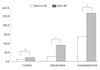
The mean serum concentrations of all stress hormones such as cortisol, epinephrine, and norepinephrine were significantly elevated in the morning after SD (Fig. 1). The reference ranges used at Samsung Medical Center are as follows: cortisol, 5.9-26.1 µg/dL; epinephrine <60 µg/dL during sitting; and norepinephrine, 120-680 µg/dL during sitting. However, mean serum concentrations of glucose and inflammatory markers (ESR and CRP) were not changed after SD. Reference ranges are as follows: ESR, 0-22 mm/h; CRP, 0-0.3 mg/dL.
Continuous Performance Test
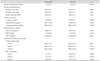
The mean score of correct responses was significantly decreased at the highest (most difficult) step after 24 h of SD; there were no statistical differences at easier steps. In contrast, the mean scores of error responses were significantly increased at all steps after 24 h of SD (Fig. 2). No significant correlations were found between CPT scores and the serum concentrations of stress hormones, glucose, and inflammatory markers. Detailed data of patients before and after SD are listed and compared in Table 1.
Discussion
The hypothesis underlying this study was that acute SD for 24 h negatively influences daytime function and increases hormone levels in healthy volunteers. As expected, the fatigue level increased and alertness decreased over time in our subjects. After a total SD of 24 h, subjects recorded a VAS score of 95 (extremely fatigued), and an SSS alertness score of 6 (foggy, losing interest in remaining awake, and slowed-down state). The CPT used in this study measures a person's sustained and selective attention and working memory. Our results showed that at easier steps (levels 1 and 2), the mean scores of correct responses were not significantly decreased after SD, but they were significantly reduced (decreased by 2.2%) at the most difficult step (level 3). The mean scores of error responses were significantly increased at all levels (increased by 69.5-136%) after SD. These findings show that attention and working memory are impaired after acute SD of 24 h, with the performance decrements being more dramatic for more complex tasks. Interestingly, subjects exhibited a relatively good performance in correct responses at easier steps after SD, but they made errors frequently even at easier steps as well as most difficult level. The findings suggest that the SD condition results in the subject being more susceptible to making an error.
Regardless of the task, cognitive performance progressively deteriorated for increasing time spent on the task; this is a classic "fatigue" effect that is exacerbated by sleep loss.9 Previous findings that performance on even brief cognitive tasks that measure attention and working memory was sensitive to SD2,10,11 support our results.
This study showed that acute (24 h) SD induces increases in the serum concentrations of stress hormones, and suggests that SD itself is the stress-eliciting situation. Although the increased concentrations of hormones after SD were still within normal ranges, statistically heightened levels of hormones after SD may indicate the important effect of sleep on stress hormones. Some studies have shown that stress hormones may interfere with memory and other cognitive functions. A significant relationship between pre-retrieval stress or high cortisol levels and impaired memory retrieval has been reported consistently.12,13 Acute elevations of exogenous glucocorticosteroids were shown to impair working memory without affecting declarative memory.13,14 A significant association was found between insomnia duration and left dorsomedial prefrontal damage in patients with focal brain lesions.15 The gray-matter volume in the left orbitofrontal cortex is reduced in chronic insomnia, and was found to be strongly correlated with the subjective severity of insomnia.16 Although the aim of the present experiment was to examine 24 h SD, significant deficits in attention and working memory after SD suggested that acute SD causes a deterioration in prefrontal function.
While cross-sectional and longitudinal studies point to a U-shaped relationship between self-reported sleep duration and the risk of type 2 diabetes mellitus,17,18 it was recently reported that prebreakfast concentrations of blood glucose and other inflammatory markers were not changed after restricting sleep (to <4.5 h/day) for 2 nights.19 We also found unchanged serum concentrations of glucose after SD. Whether or not acute SD alters glucose metabolism therefore remains uncertain. Additional measurements of other hormones such as insulin and glucagon would be helpful to clarify this issue.
Insufficient sleep and poor sleep quality are risk factors for inflammation-related conditions.19-21 Total SD for 40 h caused significant increases in E-selectin, intercellular adhesion molecule 1, interleukin-1b, and interleukin-1Ra, and decreases in CRP and interleukin-6, suggesting that sleep loss triggers a stress response that includes stimulation of both pro- and anti-inflammatory proteins.22 In contrast, we did not find any differences in the serum concentrations of ESR and CRP, which are nonspecific measures of inflammation. It is not clear whether the difference in these results is attributable to the duration of sleep deprivation or the specificity of inflammatory markers.
While this study has not revealed a direct relationship between stress hormone levels and cognitive function, we did find that acute SD for 24 h significantly heightened the levels of stress hormones and caused a deterioration of cognitive function. The acute SD condition appears to render the subject more susceptible to making an error.

 XML Download