

 PDF
PDF ePub
ePub Citation
 Print
Print


Introduction
Repetitive nerve stimulation (RNS) test is fundamentally an electrophysiologic study for the differential diagnosis of neuromuscular junction (NMJ) disorders, although there are considerable limitations in its diagnostic sensitivity and specificity. Amyotrophic lateral sclerosis (ALS) is occasionally misdiagnosed as myasthenia gravis (MG), showing prominent bulbar muscle weakness without definite atrophy of the limb muscles, or fasciculation. Although ALS is not primarily a disease of the NMJ, there have been several reports of fatigable weakness and RNS abnormalities, especially of clinically involved muscles.1-6 Significant decrements in the facial muscles such as the orbicularis oculi (OO) or nasalis (NA) muscles have only rarely been reported in ALS patients.7
The clinical significance of the RNS test was determined in this study by retrospectively analyzing the RNS tests of ALS patients with only or predominant oropharyngeal manifestations.
Methods
The data of ten patients were analyzed after the diagnosis of ALS was established. They visited the electrophysiology laboratory in the Department of Neurology at Sinchon Severance hospital with predominant oropharyngeal symptoms between January 1997 and December 2008. The serum titer of acetylcholine receptor antibody was measured in five patients. The diagnosis of ALS was confirmed by the clinical course and follow-up electrophysiologic studies.
RNS test was performed on the facial nerve with a recording electrode over the OO and NA muscles, on the spinal accessory nerve with a recording electrode over the upper trapezius muscle, and on the ulnar nerve with the recording electrode over the flexor carpi ulnaris and abductor digiti quinti muscles, using the belly-tendon method. The changes in compound muscle action potential (CMAP) amplitude followed by 3- and 5-Hz supramaximal low-rate stimulation (LRS) were analyzed. For each train of repetitive stimuli, the amplitudes of the first and fifth CMAPs were compared, and the resulting decrement of the latter (if present) is expressed as a percentage. Decrements greater than 10% were considered as abnormal according to the guidelines of American Academy of Emergency Medicine Quality Assurance committee.8
Results
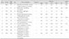
Six of the ten patients were women and their mean age was 55.3 years (range: 42-68 years). The mean duration of symptoms was 9.7 months (range: 2-36 months). All of the patients complained of only or predominant oropharyngeal symptoms including dysarthria, dysphagia and/or nasal voice. In addition to these oropharyngeal symptoms, one patient (case 1) complained of intermittent clumsiness of both hands of 1-month duration, and another (case 7) had experienced difficulty elevating the head for 1 month. None of the patients showed any clinical evidence of facial muscle weakness, ptosis or diplopia. Six patients complained of muscular fatigue or diurnal fluctuation, and two patients (cases 7 and 9) had been previously diagnosed and treated for MG (Table 1).
Abnormal decrements during 3-Hz LRS on more than one facial muscle were noted in three patients (30.0%): in two patients (cases 2 and 7) in both the OO and NA, and in one (case 9) in the OO. Borderline decrements (9.7%) during 5-Hz LRS were recorded in the NA in one other patient (case 10). None of the patients showed any abnormal decrements in the limb muscles.
Discussion
While the clinical course is very important for the differential diagnosis of ALS and MG, the pharmacologic response to anticholinesterase, the determination of anti-acetylcholine receptor antibody titer and electrophysiologic studies including the RNS test are the main ways of ensuring a differential diagnosis.
There are several reports of significant decrements on RNS test of involved limb muscles in ALS.1-6 Denys et al.2 proposed a relationship between muscle atrophy and decremental responses. However, Bernstein et al.3 reported that disease activity and speed of progression might be more important factors for these findings. Restivo et al.7 reported significant decrements in facial muscles in one ALS patient, but this case seem-ed to be a true MG patient with ALS, as evidenced by seropositivity and abnormal decrements in the deltoid muscle.
Abnormal decrements on RNS test suggest the presence of instability of neuromuscular transmission in ALS. The underlying pathogenesis of NMJ instability remains unclear, but the following hypotheses have been proposed. First, motor neuron death causes enlargement of the motor unit territory by collateral sprouting, leading to a reduced safety factor for the NMJ in re-innervated end plates.2,3,5 Second, neuronal degeneration in ALS might disturb axonal transport of the synthetic enzyme choline acetyltransferase.1,3 Third, pathologic changes occur in the NMJ in ALS, as identified in a few muscle biopsies and animal studies.2,9-11
Our results suggest that the facial muscles are involved in some ALS patients with prominent oropharyngeal symptoms, although facial weakness may not be clinically evident. It is understandable that abnormal decremental responses were not recorded in the flexor carpi ulnaris, abductor digiti quinti, and trapezius muscles, because these muscles were not involved at the time of RNS tests. We believe that our patients are not true MG cases because of their seronegativity for MG and poor response to anticholinesterase treatment, as well as their clinical courses. However, it is difficult to reach a firm conclusion because this study was retrospective and included only a small number of subjects. For example, it might be inferred that the three patients who exhibited decrement in the facial muscles presented oropharyngeal manifestations more rapidly than the others. Therefore, the speed of disease progression might be associated with neuromuscular instability in ALS, as indicated by Bernstein et al.3 Further comparative studies with larger cohorts of ALS patients are required to clarify this.
In summary, the facial muscles may be involved in ALS with predominant oropharyngeal symptoms, although facial weakness may not be observed clinically. This study makes clear that we cannot simply diagnose MG because the significant decrement of facial muscles is observed to low-rate RNS test.
 XML Download