

 PDF
PDF ePub
ePub Citation
 Print
Print


Introduction
The term "toxic leukoencephalopathy" encompasses various diseases that may injure and cause structural alteration of the brain white matter; the insults may be toxic or metabolic secondary to chemotherapy or immunosuppressive therapy, environmental, or infectious in origin.1,2 White-matter tracts devoted to higher cerebral function may be involved, causing clinical features that range from inattention, forgetfulness, and changes in personality, to dementia, coma, and death.1
Oriental medicines have been associated with severe psychiatric, neurological, and other adverse medical events. It is therefore important that psychiatrists, neurologists, and associated health-care providers are aware of such side effects. Occasionally, these medicines can cause a typical, reversible toxic encephalopathy; however, most such cases are not recognized because these adverse events are complex and are associated with other systemic signs and symptoms.3
We describe herein a married couple with progressive dementia, akinetic mutism, and toxic leukoencephalopathy, as demonstrated on brain magnetic resonance imaging (MRI), after a single dose of the same herbal medicine.
Case Report
The wife
A 61-year-old woman with a 3-year history of total thyroidectomy due to thyroid papillary cancer was admitted to hospital with behavioral changes over the previous 5 days. Fifteen days prior to her admission, the patient had taken one pack of herbs as a tonic medicine, which contained Astragalus, Angelica, Cnidium, Chrysanthemum, Aucklandia, Atractylodes, Bupleurum, and Cimicifuga species, ginger, cornus fruit, citrus peel, licorice, and other unknown adjuvants. The next morning the patient developed severe nausea, vomiting, and indigestion; these symptoms persisted for 3 days. Over a period of several days thereafter, the patient had poor oral intake, generalized aches and weakness. Five days before admission the patient developed inappropriate and incongruent speech, poor comprehension, disorientation, and repetitive meaningless behavior. One day before admission she developed akinetic mutism, lethargy, and withdrawal. There was no personal or family history of neurological diseases. The patient had worked as a teacher for an alternative school in a rural environment and there was no history of exposure to carbon monoxide.
On neurological examination the patient was alert, but she did not respond to verbal or sensory stimuli. There was no spontaneous movement during the examination. The patient's motor responses to pain were contraction and symmetric muscle spasticity. The deep-tendon reflexes were hyperactive in all extremities. There were no abnormal movements, neck stiffness, or pathological reflexes.
Laboratory studies including a complete blood count, blood chemistry, thyroid function testing, venereal disease research-laboratory testing, autoimmune vasculitis markers, HIV antibodies, vitamin B12, and folate all produced normal results. The cerebrospinal fluid had a normal opening pressure with a leukocyte count of 1/mm3, a protein level of 45.1 mg/dL, and a glucose level of 62 mg/dL. Cerebrospinal fluid smears and cultures were negative for bacteria, virus, mycobacterium, and fungus, as was the Western blot test for the 14-3-3 protein. The results of the following laboratory tests were also either normal or negative: thyroid stimulation hormone receptor antibody level, antimicrosomal antibody level, pituitary hormone level, ammonia and toxicology screening (paraquat, amphetamine, cocaine, tetrabromophenolphthalein ethyl ester, and ethanol). Hair and serum assays for the following heavy metals were either normal or negative: cadmium, cobalt, molybdenum, uranium, beryllium, sulfate, chromium, aluminum, copper, lead, mercury, selenium, arsenic, nickel, and silver. A direct assay for herbal medicine using the Cyantesmo kit did not reveal cyanide.4
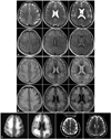
An electroencephalographic examination revealed generalized delta activity at 3-6 Hz bilaterally, but no epileptiform discharge was observed. Brain MRI revealed diffuse, symmetric, high signal intensities in both the periventricular white matter and basal ganglia medial portion in T2-weighted and fluid-attenuated inversion recovery (FLAIR) images, while diffusion-weighted, T1-weighted, and postcontrast T1-weighted images were normal (Fig. 1A). The follow-up MRI performed 1 month after admission revealed more extensive symmetrical high signal intensities in T2-weighted and FLAIR images (Fig. 1B). On the diffusion-weighted images, the lesion had a high signal intensity, with a low signal intensity on the apparent diffusion coefficient map (Fig. 1C).
The patient's mental state deteriorated rapidly and she became stuporous 1 week after admission; 1 month later her condition gradually improved and she was alert without awareness. Six months after discharge the patient was able to ambulate in a wheelchair and communicate with family members.
The husband
A 68-year-old man (the husband of case 1) was found wandering in the hospital construction site 8 days after his wife's admission. Two days previously he had become lost after patient visiting hours. He had taken the same oriental medicine as his wife, on the same day. The next morning he developed nausea and vomiting to a lesser extent. Over a period of days he developed generalized aches and weakness. There was no history of medical or neurological diseases. The patient had worked as a minister in a rural environment.
On examination the patient was alert but without awareness. Almost no meaningful speech was elicited. His motor responses to pain were contraction and symmetric muscle spasticity. His deep-tendon reflexes were hyperactive in all extremities; there were no abnormal movements, neck stiffness, or pathological reflexes.
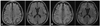
The same laboratory studies were performed as for case 1, and they revealed no abnormalities. The brain MRI revealed hyperintense lesions of the white matter bilaterally on T2-weighted, FLAIR, and diffusion-weighted images, similar to case 1 (Fig. 2A). Follow-up MRI performed 1 month after admission revealed more extensive hyperintense lesions (Fig. 2B).
The progression of this patient's neurological condition was similar to that of case 1, improving gradually after 1 month. He was able to ambulate with assistance from his family after 6 months.
Discussion
Toxic leukoencephalopathy is a disorder that is related primarily to the use of leukotoxic therapeutic agents, illicit drug use, and occupational exposure to toxins. Most patients typically present with neurobehavioral dysfunction reflecting diffuse involvement of the cerebral white matter.1 A heterogeneous array of causes has been described that can lead to toxic leukoencephalopathy with a similar clinical presentation.5
There have been few case reports of toxic encephalopathy associated with the use of oriental medicine.6,7 However, in most cases of toxic encephalopathy associated with oriental medicine, the direct toxicity of each herb and the toxic effects of a mixture of herbs have been neither verified nor reproduced. Contamination by heavy metals or toxins during the manufacturing process may also play a role.
The findings of neurobehavioral symptoms and unusual white-matter signal changes in the two cases reported herein following the ingestion of oriental medicines suggest a possible relationship, although no heavy metals or toxins were detected. It is of course possible that the toxic leukoencephalopathy is unrelated to ingestion of the oriental medicine. In addition, some unknown event or environmental exposure cannot be ruled out as the cause of this condition. However, we know of no naturally occurring illness with all of the features exhibited by this couple. We therefore believe that exposure to oriental medicines can result in toxic encephalopathies, although we have not unequivocally established a cause-and-effect relationship. Neurologists and related health-care professionals should consider oriental medicine as a possible cause in unresolved cases of toxic leukoencephalopathies.
 XML Download