

 PDF
PDF ePub
ePub Citation
 Print
Print


Introduction
Myasthenia gravis (MG), which is characterized by fatigability and fluctuating weakness of the skeletal muscles, was one of the neurological diseases with a serious prognosis in the past, as indicated by the origin of its name. MG is probably the best understood one of the autoimmune disorders of the nervous system. The main pathogenesis of MG is the loss of acetylcholine receptors (AChRs) on the postsynaptic membrane of the neuromuscular junction (NMJ) as a result of the production of AChR antibodies (Abs), although other antigens are subject to immune attack in a small number of patients.1-3 Based on the clinical manifestation, the disease is usually classified into ocular MG and generalized MG. Ocular MG affects only the extraocular muscles, whereas generalized MG affects other muscles beyond the ocular muscles, and may include limb, bulbar, facial and respiratory muscles. Serologically, AChR Abs are detectable in approximately 50% of ocular-MG cases and 80-85% of generalized-MG cases.1-3 Approximately 40% of generalized-MG patients who lack AChR Abs have been found to have Abs directed against the muscle-specific receptor tyrosine kinase (MuSK) in the postsynaptic memebrane.1-3 Patients who are negative for both AChR and MuSK Abs are now classified as "seronegative" MG.
Extensive analysis of the anti-AChR response in MG and in its experimental model, experimental autoimmune myasthenia gravis, has revealed that the autoimmune attack is dependent on T-cells, resulting from loss of tolerance toward self-antigens at the level of the thymus.1-3 However, Abs and complements are the key effectors of the loss of postsynaptic AChRs and associated destruction of the NMJ.1-3 Therefore, the goal of MG treatment is to interrupt the autoimmune process by T-cells and B-cells as soon as possible and thereby prevent further destruction of the NMJ. Since the introduction of corticosteroids (CSs) in the 1950s, immunomodulating therapies including thymectomy, intravenous immunoglobulin (IVIg), and some immunosuppressants (ISs) have been widely used. However, randomized controlled trials have been limited, perhaps because MG is a rare disease and it is difficult to recruit many proper patients. This may also be attributable to the lack of reliable and validated outcome measures. For this reason, most neurologists have chosen immunotherapies available within their medical environments in light of their own clinical experiences. The aim of this article was to review and summarize the current strategies for MG treatment and to introduce new therapeutic trials.
Symptom-Relieving Treatments
Non-selective acetylcholinesterase inhibitors
Acetylcholinesterase inhibitors (AChEIs) have been used extensively as a basic treatment and diagnostic tool for MG since 1934. Their mechanism of action is competitive blockade of the enzyme AChE, which is located in the extracellular matrix of the folded postsynaptic muscle endplate membrane and breaks down ACh into the inactive metabolites choline and acetate. AChEIs therefore prolong the level and duration of action of the neurotransmitter ACh. AChEIs are generally effective in relatively early or mild MG, in which patients have a sufficient number of remaining AChRs.2 Several AChEIs are currently available, which are generally classified according to their duration of action. The most commonly used drug is pyridostigmine, which is available in 60-mg tablets and begins to work 30 minutes after oral administration, with the action duration of 3-6 hours.1 It is generally taken every 4 hours while awake. Its dosage should be adjusted to 60-960 mg/day depending upon the clinical response and needs of the patient. The dosage is lower in patients with renal failure because it is excreted renally. Sustained-release tablets, taken at bedtime, are useful for patients with early-morning weakness, while the syrup formulation is helpful for children or patients with a nasogastric tube. AChEIs are well tolerated by most patients and are regarded as safe. Since AChEIs act on both muscarinic and nicotinic synapses, they induce the corresponding adverse cholinergic effects.1 The muscarinic effects include gastrointestinal tract hypermotility (e.g., abdominal pain and diarrhea), excessive salivation and respiratory secretions, increased sweating, and bradycardia or arrhythmia.1 The nicotinic side effects are muscular fasciculation or twitching.1 Overdose may give rise to serious cholinergic toxicity or crisis (e.g., flaccid paralysis or respiratory failure), which results from overstimulated neuromuscular transmission by excessive ACh.1
Avoidance of drugs causing disturbance of neuromuscular transmission
Some drugs can cause disturbance of neuromuscular transmission by acting on the NMJ presynaptically or postsynpatically.1 These drugs include depolarizing or nondepolarizing neuromuscular blockers, some antibiotics (e.g., aminoglycosides, macrolides, polypeptides, and monobasic amino-acid antibiotics), anticonvulsants (e.g., phenytoin and trimethadione), β-blockers, antiarrhythmic agents, calcium channel blockers, iodinated radiographic contrast agents, magnesium, phenothiazine, lithium, and chloroquine.1 In addition, some drugs are capable of inducing an anti-AChR autoimmune response de novo. Those inducing the autoimmune form of MG include D-penicillamine, interferon (IFN)-α, and pyrithioxine.1 Physicians should therefore ensure that they understand the pharmacokinetic mechanisms of these drugs and take into consideration their potential serious effects when determining treatment regimens.
Immunomodulating Treatments
Rapid induction of remission
AChEIs do not affect the production of auto-Abs and merely improve MG symptoms. The initial aim in the treatment of progressive MG should be to actively modulate the derangement of the immune system as soon as possible. Plasma exchange (PE) and IVIg have been used to induce rapid improvement of myasthenia symptoms because they work within days to 1 or 2 weeks after initiation.1 Therefore, they are useful during acute worsening of myasthenia, including myasthenic crisis and in the perioperative period. However, their effects are short-term (generally 1-2 months), and other ISs are usually necessary to maintain a state of remission.1-3
Intravenous immunoglobulin
IVIg, a blood product composed of pooled immunoglobulin G (IgG) from blood donors, has been used to treat many immune deficiencies or autoimmune disorders. IVIg acts on the immune system in various ways, including accelerating the catabolism of IgG, suppressing Ab production and neutralizing auto-Abs by anti-idiotypic Abs, inhibiting complement activation and membrane attack complex formation, and inhibiting Fc receptor function.4 The improvement rate associated with IVIg has been reported to be more than 70%.5
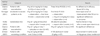
Table 1 provides a summary of randomized clinical trials involving IVIg. Wolfe et al.6 compared the efficacy of IVIg with that of a placebo in a small number of MG patients (six patients receiving IVIg and nine receiving placebo). Their randomized, open-label trial revealed a positive trend for IVIg after 6 weeks.6 One large, randomized, placebo-controlled trial conducted in patients with worsening weakness produced the promising finding that IVIg improved muscle strength (class I evidence).7 Two randomized controlled trials found no significant difference between the efficacies of IVIg and PE, although IVIg therapy was less toxic.8,9 Accordingly, it has been accepted that while their treatment efficacies are similar, IVIg is a safer option than PE. A single trial found that IVIg was superior to placebo for the treatment of MG exacerbations.7
IVIg is generally given at a total dose of 2 g/kg over 3 or 5 days. Large, randomized, double-blind trials did not show that IVIg therapy for two successive days was better than 1-day IVIg and 1-day placebo therapy.10 Its therapeutic effect appears within a couple of days from the initiation of treatment and lasts at least for several weeks, and usually up to 2 months.1 Most of the common adverse effects are mild and known to be related to the infusion process: headache, chills, myalgia, chest discomfort or shortness of breath can occur early in infusion, and are usually relieved by slowing of the infusion rate.1 Some patients exhibit skin reactions, such as urticaria or erythematous rash, which can develop up to a few days after infusion.4 In rare cases it causes more serious side effects, such as aseptic meningitis, proteinuria, or acute renal tubular necrosis in patients with underlying renal disease, and thromboembolic events including myocardial infarction, pulmonary embolism, and stroke resulting from increased plasma viscosity.1,4 Severe anaphylaxis might also occur in patients with a severe IgA deficiency.1,4
Plasma exchange
PE is an extracorporeal blood purification technique. The basic mechanism of PE is the rapid elimination from the plasma of a large amount of causative auto-Abs and other humoral mediators including cytokines and immune complexes.11 Although randomized controlled trials regarding the efficacy of PE in MG are lacking, PE is considered an established therapeutic option for preoperative preparation and MG crisis (class III evidence).11 The clinical efficacy of PE varies from 55% to 100%.12 The standard method is to remove 2-3 L of plasma three times a week or every other day for five procedures.1,3 In most patients, clinical improvement occurs after two to three procedures and usually lasts for 1-2 months.12 Adverse effects include some side effects related to the central venous catheter, such as pneumothorax, thrombosis, or catheter-related infection, and others related to the replacement fluids and anticoagulant including hypocalcemia, acid-base imbalance, and bleeding.1,12 More serious side effects include anaphylaxis and transfusion-related infection.1,12
The newly developed procedures of double-filtration plasmapheresis (DFPP) and immunoadsorption (IA) techniques have been tested with a view to reducing unwanted adverse effects and more selective removal of circulating Abs. DFPP utilizes two filters; the first filter separates plasma from blood and the second separates albumin from larger molecules including immunoglobulins, immune complexes, and lipoproteins.12 DFPP requires the instillation of less albumin solution as a replacement fluid than does classical PE, thus reducing the risk of infection.12 The IA method selectively adsorbs most large proteins such as AChR Abs by using an affinity-type adsorbent, tryptophan-linked polyvinyl alcohol gel (Immunosorba IM-TR, Asahi Medical, Japan) or a staphylococcal protein A column (Excorim, Lund, Sweden).12 Replacement fluids are not required in these methods. The clinical efficacy of these methods was reported to be similar to that of classic PE.13 Liu et al.14 recently conducted a randomized, controlled, 3-armed trial comparing DFPP (n=15) and IA (n=10) with IVIg (n=15) in 40 patients with late-onset MG. They found that both DFPP and IA exhibited better short-term clinical effectiveness than IVIg transfusion, rapidly and effectively clearing the pathogenic Abs in their patients.14 More randomized controlled trials are required to confirm these findings.
Long-term maintenance of remission
Thymectomy
Thymectomy for MG treatment has been performed widely since Blalock and his colleagues first reported its usefulness in 1939.15 Although there are no randomized controlled trials, clinical experience has led to the general acceptance that thymectomy increases the chances of remission and reduces the risk of long-term IS treatment. Thymectomy remains a generally accepted treatment for generalized-MG patients with disease onset before the age of 50 years.2 There is disagreement concerning the use of thymectomy in children prior to puberty. Some authors report favorable results with thymectomy in juvenile MG patients, whereas others have taken a more cautious attitude in this regard for very young children because of the possibility of compromising immune function.16,17 Furthermore, thymectomy is still not recommended for the treatment of anti-MuSK-Ab-positive patients because they lack pathologic thymic changes.18,19 However, there is no sound evidence supporting this. In our opinion, thymectomy should be considered first to treat generalized-MG patients with thymic abnormality with onset between the ages of puberty and around 50 years.
Many studies suggest that the earlier in the course of the illness that thymectomy is performed, the better the treatment results are, especially within the first 2 years of the disease.2,20 The therapeutic effect of thymectomy generally occurs after about 1 year, and remission is most likely in the period 5-10 years after the surgery.2,20 The reported complete remission rate after thymectomy has varied from 8.3% to 53.1%.20,21 Several procedures are used to remove the thymus, including transsternal, transcervical, transsternal-transcervical "maximal", and video-assisted thoracoscopic surgery (VATS) approaches. The standard and preferred approach has been via sternotomy, but minimally invasive VATS is becoming popular due to the associated short hospitalization, low operation-related mortality and morbidity, and cosmetic advantage. Meyer et al.22 compared the outcomes of extended transsternal thymectomy (n=47) and VATS (n=48), and found that 95.8% of the VATS group and 97.9% of the transsternal group were in complete stable remission, pharmacologic remission, or minimal manifestations state after the procedure. Meyer et al.22 concluded that these approaches resulted in equivalent outcomes. Further large, randomized studies are necessary to provide a more complete comparison of the relative therapeutic efficacies of the different thymectomy procedures.
Corticosteroids
CSs are universally used as the first-line IS for inducing comparatively rapid remission and bridging to long-term maintenance therapy using other ISs until they take effect. The mechanism of action of CSs in MG is poorly understood. The effects on the immune system involve several important pathways, including 1) reducing the distribution and trafficking of leukocytes, 2) specific inhibition of the recruitment and migration of lymphocytes to an inflammatory site, 3) blockade of various T-cell functions, 4) reducing the production and secretion of cytokines and other immune mediators from macrophages or T cells, and 5) decreasing the maturation of dendritic cells.23 The high doses generally used to induce a remission can also induce apoptosis of various immune cells.
Several studies of CSs in MG found that remission or a marked improvement occurs in an approximately 80% of cases.24 Table 2 lists the randomized clinical trials of CSs performed to date. The only adequately sized randomized controlled trial, which was conducted in 1964, compared administration of adrenocorticotrophic hormone with placebo in 43 ocular-MG patients, and found no significant difference between the two.25 That study was flawed by its short follow-up time and failure to take into account the temporary worsening of MG that occurs 1-2 weeks after initiating high-dose steroid therapy. A small randomized study of 13 severe-MG patients found no clinical difference between 100-mg prednisone (PD; 6 patients) and placebo (7 patients) administered on alternate days.26 However, that study involved only a small number of subjects and failed to provide detailed clinical data. In a double-blind, placebo-controlled study comparing IV methylprednisolone and placebo in 20 patients, the IV methylprednisolone group exhibited a significant improvement in myasthenic muscle score, while the placebo group did not (class I evidence with limited power).27
The oral administration of CSs at a dose of 1 mg/kg is commonly used for the rapid improvement of myasthenic symptoms. It is noteworthy that high-dose steroids can temporarily exacerbate myasthenia symptoms in some patients, the so-called steroid dip, which usually occurs 4-14 days after initiation and lasts for less than 1 week.1-3 Hospitalization and close monitoring for respiratory insufficiency may be needed for the prompt management of steroid-induced worsening, and IVIg or PE can be used to prevent or reduce the risk of this occurring when high-dose steroid therapy is commenced. To avoid this exacerbation, some physicians titrate up the doses of steroid slowly or administer the medication on an every-other-day schedule.
The many severe side effects of long-term use of steroids mean that this should be avoided. Common side effects are Cushing's syndrome, osteoporosis, weight gain, hyperglycemia, diabetes, hypertension, gastritis or ulcer, skin fragility, anxiety/depression/insomnia (steroid psychosis), avascular necrosis of the joints and steroid myopathy.
Long-term periodic intravenous immunoglobulin
IVIg has been used chronically when patients show an excellent response to IVIg and are contraindicated or unsuitable for ISs. In this case IVIg is administered as two or three infusions every several weeks. Wegner and Ahmed28 described a small clinical trial with long-term IVIg monotherapy in six anti-AChR-Ab-positive patients. All of the patients received an initial infusion of IVIg at a dosage of 400 mg/kg/day for 5 days followed by maintenance therapy of 400 mg/kg for 1 day every 3-4 months. After a follow-up period of 2 years, all of the patients maintained good functional status without any deterioration or IVIg-related side effects.28 If there is no subordinate restriction, including the high cost of IVIg therapy, long-term periodic IVIg may be a good option for MG patients who are unsuitable for steroid or other ISs.
Blocking the synthesis of DNA and RNA
Azathioprine
Azathioprine (AZA) is a DNA-synthesis inhibitor that inhibits the synthesis of purines (adenosine and guanine). It is a prodrug that is metabolized to several active metabolites: 6-mercaptopurine, followed by 6-thiouric acid, 6-methylmercaptopurine, and 6-thioguanine.23 These metabolites compete with the purine derivative hypoxanthine, and are incorporated into replicating DNA, consequently blocking DNA and RNA synthesis.23 AZA mainly affects the actively proliferating cells, including lymphocytes, and is thus used as one of the main drugs in organ transplantation and autoimmune disease.
Treatment of MG with AZA is associated with a good outcome, and its effectiveness has been reported to be more than 75%.29,30 Table 3 summarizes the randomized clinical studies of AZA. A few randomized trials of AZA found no significant differences between AZA and CS in improving MG.31,32 Palace et al.31 carried out a randomized, double-blind trial of AZA plus PD versus placebo plus PD in 34 generalized-MG patients, and found no significant difference between the two groups with regard to muscle strength scores and median PD dosage at 12 months. However, the median PD dose at 36 months was significantly reduced and the duration of remission was significantly longer in the AZA and PD group.31 Therefore, although this (class I) trial showed no added effectiveness of AZA in inducing remission, it did demonstrate its effectiveness as a steroid-sparing agent capable of reducing the side effects of CSs.
The initial dosage of AZA is 1 mg/kg/day with two divided doses, gradually increasing to 2-3 mg/kg/day. Its effect is generally noted 3 months after the initiation of treatment. AZA has been used widely in MG because it has fewer side effects than CSs. Common side effects are nausea, vomiting, rash, hepatotoxicity, pancreatitis, leukopenia, and thrombocytopenia.1 Therefore, regular checkup of the complete blood count (CBC), liver function, and amylase is necessary. The CBC needs to be monitored weekly during the first month, monthly for the following year, and then every 3-6 months thereafter.1 If the total white blood cell count drops to less than 3,000/µL, AZA should be discontinued for a few days and adjusted to keep the white blood cell count at more than 4,000/µL.33 It is also important to bear in mind the risk of opportunistic infection and malignancy during long-term treatment. About 10% of the general population have a deficiency of thiopurine 5-methyltransferase, which is the enzyme that blocks the formation of 6-thioguanine nucleotides, and may cause bone marrow suppression at lower doses due to the increased toxicity of its metabolites.23
Mycophenolate mofetil
Mycophenolate mofetil (MyM) is a prodrug of mycophenolic acid that inhibits inosine monophosphate dehydrogenase, which is the rate-limiting enzyme in the de novo synthesis of guanosine nucleotides. The inosine monophosphate dehydrogenase enzyme has two isoforms: type I is found in resting cells and type II is expressed by activated lymphocytes.23 The type II isoform is more sensitive to inhibition by mycophenolic acid than is the type I isoform, and hence MyM acts primarily on activated T- and B-lymphocytes.23 As a result, this drug is very specific to activated lymphocytes.
The half-life of MyM is 16-18 hours, and it is excreted renally through hepatic metabolism. The administered dosage is started at 500 mg twice a day orally and then increased to a standard dosage of 2,000-3,000 mg/day in two divided doses after 1 week. More than 50% of patients respond well to MyM treatment (as evidenced by open-label trials).34 Table 4 summarizes the randomized studies of MyM. A randomized double-blind trial of MyM plus either cyclosporine (CyA) or IS versus placebo plus either CyA or IS was conducted in 14 MG patients for 5 months.35 However, the small number of patients and short study duration meant that this study did not provide any meaningful results. Two additional randomized, double-blind studies were carried out.36,37 One study compared 2.5 g/day MyM plus 20 mg/day PD (41 patients) with placebo plus 20 mg/day PD (39 patients) in 80 IS-naïve patients with mild-to-moderate generalized, anti-AChR-Ab-positive MG.36 The other was an international, phase-III trial of 2 g/day MyM versus placebo for 36 weeks in 176 patients with anti-AChR-Ab-positive class II-IVa MG taking CSs for at least 4 weeks.37 These studies failed to demonstrate a clear benefit of MyM in permitting the tapering of the dosage of PD.37 However, a retrospective study recently reviewed outcomes and PD dosage for 102 anti-AChR-Ab-positive MG patients with MyM alone or with PD.38 In this retrospective study, the percentage of patients with a desirable outcome began to increase after 6 months, and 80% of those followed for more than 24 months had a desirable outcome.38 After 25 months, 54.5% of patients were able to discontinue PD.38 The results of this study suggest that the clinical efficacy of MyM begins to appear after 6 months when administered as monotherapy or in combination therapy with PD (class IV evidence).38
Common adverse effects of MyM are gastrointestinal symptoms including nausea, vomiting, diarrhea and abdominal cramps, headache, fatigue, skin reactions, and viral infections. Rarely reported side effects are pure red cell aplasia, progressive multifocal leukoencephalopathy, and malignancy.39,40 CBCs should be monitored regularly during MyM treatment. MyM is contraindicated in pregnant women or women planning to conceive due to the risk of miscarriage or congenital malformations.
Cyclophosphamide
Cyclophosphamide (CP), a well-known antineoplastic agent, is a prodrug that is converted into an active metabolite (4-hydroxycyclophosphamide) in the liver. It is one of the classic alkylating agents, adding an alkyl group to the guanine base of DNA, leading to cell death.23 Perez et al.41 followed up 42 MG patients who received a total dose of 7.6-130 g of CP over 2-37 months.According to their definition (being asymptomatic for at least 6 months without medication), remission was observed in 12 of the 16 patients (75%) who were followed for more than 18 months.41
A randomized, double-blind trial was conducted in 23 MG patients with either poor disease control or steroid-related side effects, who were given either IV CP plus PD (n=12) or placebo plus PD (n=11).42 CP pulses were given at an initial dose of 500 mg/m2 of body surface, and then monthly for the first 6 months, and finally every other month thereafter.42 PD was tapered off by 10 mg/month if initially taking more than 50 mg/day, 5 mg/month if taking 20-50 mg/day, and 2 mg/month if taking 20 mg/day or less.42 Significant improvement of muscle strength was noticed in the CP group at 12 months, mainly in the bulbar, masticatory and extraocular muscles.42 A significant reduction in PD dosage was also reported in the CP group at 6 and 12 months compared with the placebo group.42
Drachman et al.43 performed a trial called "rebooting the immune system" involving high-dose CP for treatment of refractory MG patients. They followed 12 refractory patients who received 200 mg/kg of CP for 1-9 years.43 Eleven of the 12 patients exhibited a dramatic clinical improvement from 5 months to 7.5 years, and this improvement lasted for more than 1 year in 7 patients.43 They suggested that high-dose CP resulted in effective remission in most refractory MG patients.43 Therefore, this method could be considered cautiously in severe-MG patients who are refractory to other ISs. The administered dose is 3-5 mg/kg orally or 200 mg IV for 5 days. Adverse effects of CP include nausea, vomiting, diarrhea, alopecia, darkening of the skin and nails, infertility, myelosuppression, oliguria, hemorrhagic cystitis (prevented by large fluid intake and Mesna), opportunistic infection, and malignancy such as bladder cancer.23,33
Methotrexate
Methotrexate (MTX), an antimetabolite and antifolate drug, inhibits the action of dihydrofolate reductase catalyzing the conversion of dihydrofolate to active tetrahydrofolate. Folic acid and folate are necessary for purine and pyrimidine synthesis. Therefore, MTX ultimately blocks the synthesis of DNA and RNA, and has a more potent effect on rapidly dividing cells.23 Side effects associated with MTX therapy are gastrointestinal symptoms, myelosuppression, mucositis, cystitis, liver fibrosis, cirrhosis, and lung fibrosis. Combined administration with folate can reduce the risk of myelosuppression.
There have been no published clinical trials for MTX therapy in MG patients. MTX is recommended by some experts as a second-line IS in other-drug-resistant MG patients.23
Targeting T-cells: calcineurin inhibitors (cyclosporine A and tacrolimus)
Cyclosporine A (CyA) and tacrolimus (FK506) are calcineurin inhibitors that bind to the cytosolic proteins, cyclophilins (immunophilins), of lymphocytes: CyA binds to cyclophilin and FK506 binds to FK506-binding protein.23,33 Activation of T-cell receptors by antigens increases intracellular calcium and induces the activation of calcineurin.23,33 Activated calcineurin plays an important role in the transcription of proinflammatory cytokines including interleukin (IL)-2, IFN-γ, or tumor necrosis factor alpha (TNF-α) by dephosphorylating the transcription factor, nuclear factor of activated T-cells.23 The CyA-cyclophilin and FK506-binding protein complexes block calcineurin, consequently inhibiting T-cell activation and proliferation.23
Until now, uncontrolled trials of CyA were conducted in severe-MG patients who were unresponsive to thymectomy, steroids, or AZA. About 80% of recruited patients improved after CyA treatment.44,45 The first randomized, placebo-controlled trial showed that the increase in muscle strength was significantly greater in the CyA group (n=10) than in the placebo group (n=10) at 6 and 12 months.46 Tindall et al.47 followed 39 generalized-MG patients with CyA plus PD versus placebo plus PD. They showed that muscle strength was significantly increased in the CyA group, but that there was no definite difference in the percentage change in the steroid dosage between the two groups at 6 months.47
CyA administration begins with two divided dosages of 4-6 mg/kg/day, and can be adjusted to maintain the CyA blood level at 100-150 ng/mL after 1 month.33 Regular check-up for levels of blood urea nitrogen and creatinine (Cr) are required, and Cr levels should be maintained at less than 150% of the baseline value (i.e., before treatment).33 Adverse reactions of CyA include gingival hyperplasia, convulsion, nephrotoxicity, hepatotoxicity, peptic ulcer, pancreatitis, hypertrichosis, hypertension, opportunistic infection, malignancy (especially skin cancer), and posterior reversible encephalopathy syndrome.23,33,48
The use of FK506 is supported by only a few non-randomized and non-controlled studies. Nine (47%) of 19 generalized-MG patients in an open clinical trial exhibited improvement of MG or activities of daily living scores with FK506 at a dosage of 3-6 mg/day, and reduction of anti-AChR Ab titers and IL-2 production at the end of a 16-week period.49 Ponseti et al.50 reported the results of a low-dosage FK506 (0.1 mg/kg/day) trial in 212 MG patients: 110 thymectomized, CyA-, and PD-dependent patients; 68 thymectomized patients who started FK506 and PD early postoperatively (24 hours after surgery); and 34 patients older than 60 years with nonthymomatous generalized MG or in whom thymectomy was contraindicated. Of the entire cohort, 13.7% achieved clinically stable remission and 73.8% achieved pharmacologic remission.50 In particular, the results were best in subjects who took PD and started FK506 early after thymectomy.50 Furthermore, PD administration was withdrawn in 95.1% of all patients.50 Fk506 is administered orally in two divided dosages at 0.05-0.1 mg/kg and adjusted to maintain blood levels at 5-15 ng/mL.33 FK506 is less nephrotoxic than CyA. FK506 may be carefully considered for intractable steroid-dependent MG patients.
Targeting B-cells: monoclonal antibody (rituximab)
Rituximab (RTM) is a chimeric monoclonal Ab against CD20 antigen on surface pre-B and mature B-cells.23 It was initially used to treat B-cell malignancies, such as non-Hodgkin's lymphoma and large B-cell lymphoma, but its use has been expanded to include autoimmune diseases including rheumatoid arthritis, systemic lupus erythematosus, and multiple sclerosis.23
There have been only few, small, non-controlled clinical trials for RTM therapy in refractory MG patients.51,52 Zebardast et al.51 reported promising results for RTM in six refractory MG patients with anti-AChR Abs or anti-MuSK Abs, commenting that all patients showed clinical improvement with a reduced need for multiple and/or high-dose immunotherapy. Maddison et al.52 recently treated ten patients with generalized MG and two with Lambert-Eaton myasthenic syndrome with RTM. One-quarter of the patients achieved remission and 42% improved clinically over 18 months, and hence RTM may be considered as an option for refractory patients who are uncontrolled with multiple ISs.
RTM is administered intravenously weekly for 2-4 weeks at a dose of 375 mg/m2, as it is used in hematologic malignancies. Common side effects are related to the IV infusion: chills, fever, nausea, and sometimes bronchospasm. Other side effects are hepatitis B reactivation and cardiac arrhythmia.51 There have been some reports of progressive multifocal leukoencephalopathy in patients taking monoclonal Ab including RTM.53 This serious side effect renders it necessary to pay RTM therapy special attention.
New therapeutic trials
Antisense treatment (EN101)
Mammalian AChE has three isoforms: synaptic (AChE-S), erythropoietic, and read-through (AChE-R).54 AChE-S is bound to the muscle basement membrane and is responsible for the rapid hydrolysis of ACh at the neuromuscular synapse. AChE-R, which plays a role in nonsynaptic ACh hydrolysis and in morphogenesis, is present in much lower amounts but accumulates in response to stress.54 Chronic AChE inactivation by nonselective AChEIs induces the overexpression of AChE-R.54 Nonsynaptic ACh hydrolysis by overexpressed AChE-R may result in myopathic changes and limit the duration of AChEI to recover stable compound motor action potentials and anti-inflammatory responses through cholinergic up-regulation.54
EN101 (Monarsen) is a synthetic antisense RNA molecule that selectively lowers the levels of AChE-R in both blood and muscle by inhibiting the gene expression of AChE-R at the mRNA level. EN101 therapy may thus improve muscle strength.54,55
A phase-Ib open-label clinical trial with oral EN101 was recently undertaken in 16 MG patients, of which 14 exhibited a clinical improvement without serious adverse effects.55 This promising result has led to a phase-II trial for this therapy.
Tumor-necrosis-factor-alpha receptor blocker (Etanercept)
Etanercept, which is an engineered fusion protein linked to Fc of IgG1, binds to TNF.56 TNFα is a typical proinflammatory cytokine produced by macrophages and monocytes. Etanercept blocks the activity of TNFα by acting as a decoy receptor binding to TNF.56 Some clinical trials have been reported in other autoimmune diseases including rheumatoid arthritis, Wegener's granulomatosis, and ankylosing spondylitis. Etanercept therapy for MG is still in its infancy. Rowin et al.56 performed an open-label trial of 25-mg etanercept twice weekly in 11 steroid-dependent MG patients, but found that only 6 patients showed improvement after 6 months.
Etanercept may be administered as a subcutaneous injection at a dose of 25 mg twice weekly or 50 mg once weekly. It has been reported to cause neuromuscular complications, including autoimmune peripheral nerve disorders such as chronic inflammatory demyelinating polyradiculoneuropathy, Guillain-Barré syndrome, mononeuritis and MG.57 Further well-organized studies are thus required to determine the efficacy and adverse effects of this drug in MG.
Experimental immunotherapies
The complement system, which is a part of the innate immune system, consists of classical, alternative, and mannose-binding lectin pathways that activate each other.58 The complement system plays an important role in host defense against bacteria and viruses, interfacing between innate and adaptive immunity, leading to clearance of immune complexes and apoptotic cells.58 Since MG is fundamentally an autoimmune disease mediated by auto-Abs, complement plays an important role in the Ab-mediated destruction of neuromuscular synapses. A recent study of the animal model of MG, experimental autoimmune myasthenia gravis, yielded promising results for the efficacy of the C5 complement inhibitor, rEV576.59 The complement inhibitor might thus be expected to be a new therapeutic choice in MG.
Other experimental immunotherapies are being investigated, including adoptive cellular gene therapy, genetically engineered antigen-presenting cells, syngeneic AChR fragments, and preventive strategies using RNA aptamer.
Strober et al.60 recently reported the case of a 17-year-old boy with severe, refractory MG. He was previously undercontrolled with IVIg, CSs, thymectomy, AZA, MyM, plasmapheresis, RTM, and high-dose CP. He was treated with allogenic hematopoietic stem cell transplantation, and his weakness completely resolved at 40 months posttransplantation.60
Conclusions and Recommendations
The treatment of MG should be tailored according to the clinical manifestations or subtypes, functional impairment, medical state, and activities of daily living of individual patients. As mentioned above, in progressive MG it is desirable to block the abnormal immunologic process as soon as possible while minimizing the adverse reactions of therapy.
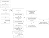
Fig. 1 summarizes our recommendations. Thymectomy may be considered preferentially in postpubertal generalized-MG patients with a stable medical condition, if thymoma is discovered on chest computed tomography. Symptomatic treatment with AChEI might be sufficient in ocular-MG patients. In uncontrolled cases with sufficiently large amounts of AChEI, steroids might be considered. If the response to steroids is incomplete, a first-line IS, such as AZA or MyM, may be added.
If generalized-MG patients has moderate to severe symptoms, IV high-dose steroids may be recommended for rapid induction of remission. Simultaneous IVIg infusion or hospitalization would be required due to the risk of transient aggravation during the early days after steroid administration. The high dose should be maintained until remission or minimal manifestations are attained, and then slowly tapered to the minimal effective dose. It is advisable to start calcium supplements and vitamin D and possibly bisphosphonates with steroid administration. Once adequate reduction of steroids is obtained, first-line IS (AZA or MyM) may be added with the purpose of steroid-sparing. If myasthenic symptoms are aggravated during tapering steroids, dose reduction should first be suspended, holding the dose steady for a few days. If the symptoms progress continuously or fail to improve, it will be necessary to increase the CS dose considerably, at which time a slight change would not help to secure a remission. An alternative is to treat the patient with a course of IVIg, which may result in a long-term return to remission. Once a remission is achieved again, tapering is retried more slowly than before and, if the patient has not been on ISs before, starting this form of treatment should also be carefully considered.
Current therapeutic modalities for MG have reduced the associated mortality and morbidity dramatically. Although advances in critical-care medicine have taken the lead in this progression, immunomodulating therapies have accelerated the improvement. CSs are the mainstay of immunotherapies for MG, but the side effects are considerable. Similarly, other ISs have many potentially serious side effects. Therefore, the development of more effective and safer ISs is a pressing issue. Various ISs that act on the immune system more specifically are being studied. The ideal model might be immunotherapy that can control selectively the AChR-specific T-cell immune response of MG.


 XML Download