

 PDF
PDF ePub
ePub Citation
 Print
Print


Introduction
Benign paroxysmal positional vertigo (BPPV) is one of the most common disorders causing dizziness.1,2 Up to 90% of positioning vertigo/nystagmus cases are attributable to BPPV,3 which has been recognized as a clinical entity since the late 1800s and early 1900s.4 In 1921, Barany described the characteristic nystagmus and vertigo induced by position change and attributed these symptoms to a disorder of the otolithic organ.5 In 1952, Dix and Hallpike described torsional vertical nystagmus provoked by a specific ear-down position with a latency of several seconds, in which the nystagmus lasted only for a limited time (usually less than 20 seconds) and the direction of the nystagmus reversed on resuming the upright position. The nystagmus also showed a fatigability with a progressive decline of intensity on repetition of the maneuvers.6 These authors coined the term "benign paroxysmal positional vertigo", and the provocative positional testing was named in their honor.
Schucknecht was the first to provide a pathophysiological concept of BPPV. In 1969 he proposed the theory of "cupulolithiasis" on the basis of pathological studies that demonstrated otolithic debris attached to the cupula.7 According to the theory, the cupula, which became heavy due to attached otolithic debris, could be deflected by position changes, thus evoking nystagmus.7 However, the concept of cupulolithiasis has several limitations and is thus unable to explain all of the characteristics of nystagmus and vertigo in BPPV. In 1979, Hall proposed the concept of "canalolithiasis", which states that otolithic debris from the utricular macule migrates into the semicircular canal via the nonampullary portion, causing vertigo and nystagmus by moving freely inside the semicircular canal and inducing endolymph flow during positional changes.8 The concept of canalolithiasis was supported by the intraoperative observation of abundant free-floating debris in the endolymph of the posterior semicircular canal.9 This concept formed the theoretical basis of the canalith repositioning maneuvers (CRMs) to treat BPPV.10,11
Prior to the 1980s it was believed that BPPV developed only in the posterior semicircular canal. However, in 1985 the concept and clinical features of BPPV involving the horizontal semicircular canal (HC-BPPV) was introduced by McClure, who described seven cases with geotropic nystagmus (nystagmus beating toward the ground) during the Dix-Hallpike maneuver without any evidence of central lesions.12 Later, the apogeotropic type of BPPV was also reported, in which the nystagmus beats toward the ceiling during lateral head turning in the supine position.13
Symptoms of BPPV
The main symptom of BPPV is vertigo (spinning sensation) induced by a change in head position with respect to gravity. Patients typically develop vertigo when getting out of bed, rolling over in bed, tilting their head back, for example to look up shelves, or bending forward, for example when fastening their shoes. However, the symptoms of BPPV may vary among patients, and may manifest with nonspecific dizziness, postural instability, lightheadedness, and nausea.16,17 The vertigo in BPPV is typically intermittent and positioning dependent. Patients with BPPV do not experience severe vertigo during usual daytime activities performed with an upright posture, but rather when they get out of bed.
The vertigo is mostly transient in BPPV, its duration correlating well with the duration of the positioning nystagmus, which usually resolves within 30 seconds in posterior canal BPPV (PC-BPPV).6 However, the duration is relatively longer (sometimes lasting longer than 1 minute) in HC-BPPV.18 Even though patients with BPPV occasionally report persistent dizziness and imbalance, careful history taking reveals that in most cases, aggravation of their symptoms occurs with position change.
Epidemiology
There is a dearth of epidemiologic studies on BPPV. The prevalence of BPPV has been reported as 10. 7-64 per 100,000 population.19 According to a recent study in Germany in which utilized telephone-based interviewing, the lifetime prevalence of BPPV is 2.4%, with a 1-year-incidence of 0.6%.20
Idiopathic BPPV is more prevalent in the elderly and in women,20,21 with a women-to-men ratio of 2-3:1 and a peak age at onset in the sixth decade of life.21 BPPV is more likely to involve the right ear, a factor that may be related to the habit of sleeping on the right side in the general population.22
BPPV mostly develops in the posterior and horizontal semicircular canals.23 PC-BPPV has been said to account for 60-90% of all BPPV cases, and HC-BPPV for 5-30% of the cases.23,24 However, HC-BPPV now appears to be more prevalent than was previously thought.23-25 The proportion of HC-BPPV-attributable cases decreases with increasing mean time interval from the symptom onset to diagnosis,24,26 probably due to the higher rate of spontaneous resolution in HC-BPPV. Thus, the relative proportion of each type of BPPV may depend upon the setting of each clinic. BPPV rarely involves the anterior semicircular canal.14 BPPV arising from multiple canals has also been described.24,27
Causes
The cause of BPPV is mostly unknown (idiopathic). In view of the high prevalence of BPPV in middle-aged women, hormonal factors may play a role in the development of BPPV.28 In a recent study, bone mineral density score was decreased in both women and men with idiopathic BPPV compared with that in normal controls without a history of dizziness.29 The prevalence rates of osteopenia (-2.5<T-score<-1.0) and osteoporosis (T-score ≤-2.5) were also found to be higher in both women and men with BPPV than in normal controls. Furthermore, in women aged ≥45 years, the lowest T-scores were also decreased in the recurrent group, compared with those in the de novo group. These findings suggest the involvement of deranged calcium metabolism in idiopathic BPPV and a significant association between osteopenia/osteoporosis and idiopathic BPPV. Otoconia are deposits of calcium carbonate in the form of composite calcite crystals, and bone contains 99% of the calcium found in the body.29 Decreased estrogen levels may disturb the internal structure of the otoconia or their interconnections and attachments to the gelatinous matrix. Alternatively, an increase in the concentration of free calcium in the endolymph due to increased calcium resorption may reduce the capacity to dissolve the dislodged otoconia.28
BPPV may develop secondary to various disorders that damage the inner ear and detach the otolith from the utricular macule.21,30-32 Head trauma causing mechanical damage to the ear is the most common cause of BPPV.32 Patients rarely develop BPPV after mastoid surgery or if they engage in a persistent head-tilt position, such as among barbers or dentists.33,34
Compared with the idiopathic form, traumatic BPPV exhibits several distinctive characteristics, including a higher incidence of bilaterality, involvement of multiple canals on the same side, equal occurrence among women and men, a younger and more even age distribution, more difficult to treat, and frequent recurrences.31,32 In addition, BPPV may develop secondary to any of the inner ear diseases (e.g., vestibular neuritis, labyrinthitis, and Meniere's disease) that give rise to degeneration and detachment of the otoconia, but do not totally impair semicircular canal function.30 The incidence of BPPV is also known to be higher in patients who suffer from migraine, even though the exact mechanism remains to be elucidated.35 BPPV has been reported to occur in association with giant-cell arteritis, diabetes, and hyperuricemia.36-38
Pathomechanism
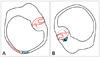
The detached otolith debris could be either attached to the cupula (cupulolithiasis) or may be free-floating in the semicircular canals (canalolithiasis) (Fig. 1). Pathological studies have shown that both of these conditions exist.7 The otolithic debris deflects the cupula and gives rise to a spinning sensation via a direct gravitational effect on the cupula or by inducing endolymph flow during head motion in the direction of gravity (Fig. 2). According to the cupulolithiasis theory, a cupular deposit (heavy cupula) would induce a gravitational effect on the crista. However, the action of free-floating debris is the currently accepted pathophysiologic mechanism of typical BPPV. According to the canalolithiasis theory, the free-floating particles move under the influence of gravity when changing the position of the canal in the earth-vertical plane. The hydrodynamic drag of the particles induces endolymphatic flow, resulting in cupular displacement and leading to the observed typical responses.9
Several studies have attempted to determine utricular (otolithic) abnormalities in BPPV, but they have produced inconsistent results.39-42 Patients with BPPV may exhibit abnormalities in vestibular evoked myogenic potentials, subjective visual horizontal, and gain during off-vertical axis rotation.40-42
Diagnosis
Each type of BPPV is diagnosed by observing the patterns of nystagmus induced during positioning maneuvers that have been designed to move only the involved canal in the direction of maximal gravity. However, accurate observations of the nystagmus require the fixation to be removed during the maneuvers.
PC-BPPV
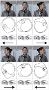
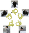
In PC-BPPV, the positioning nystagmus is typically induced by Dix-Hallpike maneuvers in the direction of the involved canal (Fig. 3).6,21 During the Dix-Hallpike maneuver, it is thought that the free-floating otolithic debris (canalolithiasis) in the posterior canal moves away from the cupula and stimulates the posterior canal by inducing ampullofugal flow of the endolymph (Ewald's first law). Excitation of the posterior canal in turn activates the ipsilateral superior oblique and contralateral inferior rectus muscles, which results in tonic downward deviation of the eyes with a torsion in the direction of the uppermost ear. Accordingly, the resultant nystagmus would be upbeating and torsional, with the upper pole of the eyes beating toward the lowermost ear. The nystagmus usually develops with a brief latency of several seconds, resolves within 1 minute (usually within 30 seconds), and its direction is reversed on sitting.6 The nystagmus diminishes (i.e., it fatigues) with repeated examinations.6 Cupulolithiasis may exist in the posterior canal. Compared with canalolithiasis, the cupulolithiatic type of PC-BPPV tends to have shorter latency and longer time constant (i.e., it is more persistent).43
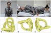
The Dix-Hallpike maneuver has been considered the gold standard for diagnosing PC-BPPV. However, this maneuver should be performed with caution in patients with a history of neck surgery, cervical radiculopathy, and vascular dissection syndrome, since it requires rotation and extension of the neck during the positioning.44 The side-lying test may be used as an alternative when the Dix-Hallpike maneuver is inapplicable; after seating the patient on the side of an examination couch, the patient quickly lies down on the side with the head turned to 45° in the opposite direction (Fig. 4).44
HC-BPPV
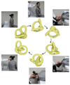
HC-BPPV is diagnosed by the supine roll test (the Pagnini-McClure maneuver), in which the head is turned by about 90° to each side while supine (Fig. 5). During this maneuver, horizontal nystagmus may beat toward the ground (geotropic nystagmus)(Fig. 5A) or toward the ceiling (apogeotropic nystagmus)(Fig. 5B). The induced nystagmus tends to be more persistent in HC-BPPV than in PC-BPPV. The nystagmus evoked during positioning in HC-BPPV usually exhibits less fatigability and a shorter latency than that evoked in PC-BPPV.18
Determination of the involved side (lateralization) is very important for the proper treatment of HC-BPPV using CRMs, which is discussed later (Table 1). Since ampullopetal flow of the endolymph evokes a greater response than ampullofugal flow in the horizontal canal (Ewald's second law), the induced nystagmus is stronger when the head is turned toward the affected ear in the geotropic type of HC-BPPV. In contrast, head turning to the healthy ear generates a stronger nystagmus in apogeotropic HC-BPPV.45-48 Determination of the involved ear is sometimes difficult due to rather symmetrical responses, especially if the induced nystagmus is not recorded. In these instances, other findings may provide clues toward determining the affected ear. In HC-BPPV, nystagmus may be induced by lying supine from the sitting position [lying-down nystagmus (LDN)] or by bending the head forward while sitting [head-bending nystagmus (HBN)].46-48 In up to 80% of HC-BPPV cases, LDN and HBN are in the opposite direction. In geotropic HC-BPPV, HBN beats mostly toward the affected ear, while LDN is directed mostly toward the healthy ear.46-48 HBN in geotropic HC-BPPV is ascribed to ampullopetal migration of the otoliths, while LDN is explained by ampullofugal displacement of the otoliths in the horizontal canal. In contrast, HBN is mostly contralesional and LDN is usually ipsilesional when observed in apogeotropic HC-BPPV.48 HBN and LDN in apogeotropic HC-BPPV are explained by deflection of the heavy cupula in response to the positional change.
In apogeotropic HC-BPPV, the induced horizontal nystagmus may disappear when the head is turned to the affected ear by 10-20°, while supine (null point).49 The null point is explained by alignment of the heavy cupula in the direction of the gravitational vector.
Spontaneous nystagmus, also known as pseudospontaneous nystagmus,50 is not uncommon in HC-BPPV. In previous reports, 66-76% of HC-BPPV patients exhibited spontaneous nystagmus.45,50 The spontaneous nystagmus in HC-BPPV may be related to the anatomical position of the horizontal semicircular canal, which is inclined 30° backwards from the horizontal plane.50 Accordingly, the gravitational force may affect the otolithic debris inside the canal or the heavy cupula, even when in the upright sitting position. For the same reason, pseudospontaneous nystagmus disappears when the patient's head is bent forwards by about 30°. In this position, since the horizontal canal is aligned with respect to the earth-horizontal plane, the effect of gravity is negated.49,50 However, pseudospontaneous nystagmus should be differentiated from continuous nystagmus with sustained vertigo resulting from so-called canalith jam and negative endolymph pressure between the plug and the cupula.51
In BPPV, spontaneous reversal of the initial positioning nystagmus rarely occurs without further position changes. In geotropic HC-BPPV, the initial geotropic nystagmus occasionally reverses its direction when the head is turned toward the lesion side, and the induced nystagmus is intense (maximal slow phase velocity=104±62°/second, mean±SD).52 Short-term adaptation of the vestibulo-ocular reflex seems to be the main mechanism underlying this spontaneous reversal of the initial positioning nystagmus.25,52
AC-BPPV
BPPV rarely involves the anterior semicircular canal, and AC-BPPV exhibits several characteristics that contrast with those of PC-BPPV. In AC-BPPV, SHH as well as the Dix-Hallpike maneuver on either side may evoke downbeat nystagmus with an ipsitorsional (upper poles of the eyes beating toward the involved ear) component.53,54 Furthermore, the torsional nystagmus in AC-BPPV may not be evident, as it is in PC-BPPV.
Mixed-canal type of BPPV
BPPV may involve multiple semicircular canals. The mixed-canal type of BPPV, the most common of which is a combination of PC- and HC-BPPV,24 comprises about 1.5-5.0% of all BPPV cases in the literature.24,55 The mixed-canal type of BPPV frequently involves the canals on the same side (e.g., right horizontal and right posterior canals),27,55 but bilateral involvement has also been reported.18 Trauma may increase the risk of mixed-canal BPPV.55
Differential Diagnosis
Most BPPV patients worry that they may be suffering from a serious disorder, such as stroke. Even though BPPV is a benign and self-remitting disease, more serious disorders such as posterior circulation stroke can mimic BPPV.56 However, central positional nystagmus usually accompanies persistent vertigo, profound imbalance, and other neurological symptoms and signs (Table 2).57
Since positional downbeat nystagmus is a typical finding in lesions involving the cerebellum,58 and AC-BPPV is a rare condition, accounting for only 1.5-5% of all BPPV cases, a diagnosis of AC-BPPV should be reserved only for typical cases without other neurological deficits.24,58 Even in those patients, the possibility of central pathology should be investigated when repeated CRMs fail to resolve the symptoms and nystagmus.
In a previous study, 72% of patients with positional downbeat nystagmus exhibited central disorders, while 24% (most of which were believed to be AC-BPPV) had unknown etiology without a central pathology.58
Treatments
BPPV is usually a self-remitting disorder and may resolve as time goes on without specific treatment. According to a report on the natural course of untreated BPPV, most HC-BPPVs resolve within 16±19 days and PC-BPPVs within 39±47 days of their onset.26 However, a correct diagnosis and proper repositioning maneuvers may allow a rapid and simple cure for the BPPV.61
By the 1970s, the treatment of BPPVs mostly involved the administration of vestibular suppressants and restriction of the position change responsible for the vertiginous spells, which meant that resolution of the condition was largely time consuming. After the Brandt-Daroff exercise was introduced in 1980, it was usually recommended that patients with BPPV perform active exercises.62 By adopting the Brandt-Daroff exercise, the treatment period of BPPV was shortened by 10-14 days. However, the aim of this exercise was habituation and compensation of the vestibular system.62 Patients with BPPV were not treated effectively until the late 1980s and early 1990s, when CRMs were introduced.
CRMs
PC-BPPV
The most popular methods for treating PC-BPPV are Semont's liberatory and Epley's maneuvers.10,11 These maneuvers employ stepwise changes in head position (Fig. 6) to flush free-floating otolithic debris out of the semicircular canals and back into the utricle. The original version of Epley's maneuver has been modified by other physicians for ease and simplicity. The mastoid vibration originally applied during the repositioning maneuvers by Epley is no longer recommended.63,64 Initially, patients were instructed to maintain an upright position for 48 hours after the repositioning maneuver; however, such position restriction after treatment is not mandatory and can be simplified.65
Theoretically, positional vertigo may be resolved after repositioning the otolithic debris into the utricle with Epley's maneuver. The reported success rate is about 80% after one round of the repositioning maneuver; the success rate increases with repetition of this protocol. According to a recent meta-analysis of the modified Epley's maneuver for PC-BPPV, the treatment demonstrated a symptom improvement rate four times greater, and a nystagmus resolution rate five times greater than the placebo group.66
The results of Epley's maneuver can be predicted even during the maneuver. When the head is turned 90° toward the unaffected side after the Dix-Hallpike maneuver, the positioning nystagmus develops in the same direction as the maneuver (orthotropic nystagmus) if a clump of particulate matter moves in the correct direction into the common crus, resulting in a successful repositioning. However, the direction of the nystagmus would reverse if a heavy cupula with attached otolithic debris deflects ampullopetally or if the particles move back toward the cupula, which implies that the repositioning will be unsuccessful.67 Epley's maneuver is the only recommended method of treating PC-BPPV, with confirmed evidence level A according to the American Academy of Neurology.68
Semont's liberatory maneuver is also helpful in treating PC-BPPV (Fig. 7),69 and may be considered an alternative treatment modality for this condition, especially in patients who have difficulty extending the neck due to spinal disorders. However, the efficacy of Semont's liberatory maneuver has yet to be established unequivocally.68 Recently, a protocol for the self-treatment for PC-BPPV has been introduced based on Epley's and Semont's maneuvers.70,71 After diagnosis by a physician, a treatment maneuver is described to the patient that can be easily performed away from the hospital.
Geotropic HC-BPPV
Rotations of 270° or 360° around the yaw axis (the so-called barbecue maneuver) toward the unaffected ear are popular methods for the treatment of geotropic HC-BPPV.72 These maneuvers consist of sequential head turning of 90° toward the healthy side while supine (Fig. 8). With these maneuvers, the free-floating otoconial debris migrates in the ampullofugal direction, finally entering the utricle through the nonampullated end of the horizontal canal.
Lying with the healthy ear downward for approximately 12 hours (forced prolonged position) can be employed, especially in patients suffering from severe symptoms who cannot perform sequential position changes.73 The Gufoni maneuver is another alternative.74-76 After being seated on an examination couch, the patient lies down on the healthy lateral side with a quick lateral movement and is maintained in this position for 1-2 minutes until resolution of the evoked nystagmus. A quick 45° rotation of the head toward the floor is then performed, with the patient maintaining this position for another 2 minutes, followed by a slow return back to the starting position. A major advantage of the Gufoni maneuver is its simplicity.
Apogeotropic HC-BPV
Apogeotropic HC-BPPV is attributed to either cupulolithiasis or canalolithiasis within the anterior arm of the horizontal semicircular canal.13,25 These explanations are consistent with the characteristics of the positioning nystagmus observed in HC-BPPV. In apogeotropic HC-BPPV, the therapeutic goal should be to detach the otolithic debris from the cupula or shift the debris from the anterior into the posterior arm of the horizontal canal.77
If the otolithic debris is attached at the utricular side of cupula, its detachment should result in immediate resolution of the positional vertigo and nystagmus. In the case of adhesion from the canal side of the cupula or free-floating particles in the anterior arm, detachment and shifting of the otolithic debris into the posterior arm would give rise to a transition into geotropic HC-BPPV.78 Therapeutic head-shaking in the horizontal plane,73,77 a modified Semont maneuver,79 and the Gufoni method80 have been proposed as treatment regimens for apogeotropic HC-BPPV.
The aim of head-shaking is to detach the otolithic debris from the cupula, irrespective of the side to which it is attached, using alternate accelerating and decelerating power.77
The modified Semont maneuver comprises the following three steps:79 1) the patient is brought briskly into a side-lying position with the affected ear downward; 2) the patient's head is turned 45° downward, with this position being maintained for 2-3 min; and 3) the patient resumes the original sitting position. This maneuver was initially designed to dislodge the debris attached to the utricular side of the cupula.77,80
In the Gufoni maneuver for apogeotropic HC-BPPV, the patient sits with the head directed straight ahead and then quickly moves into a side-lying position on the affected side, remaining in this position for 1 or 2 more minutes after the end of apogeotropic nystagmus. The head is then turned 45° upward very quickly and is kept in this position for 2 minutes, followed by a slow return to the sitting position.75,80 The Gufoni maneuver was designed to remove the otolithic debris from the anterior arm of the horizontal semicircular canal near the cupula.
AC-BPPV
Various repositioning maneuvers have also been advanced to treat AC-BPPV. In the reverse Epley maneuver, the patient submits to the same sequence of positional changes after the Dix-Hallpike maneuver on the side of the healthy ear.14 Modified repositioning maneuvers and forced prolonged position have also been adopted in treating this particular BPPV.81,82
Rehabilitation
Irrespective of the involved canals, the Brandt-Daroff exercise may be attempted when the repositioning maneuvers fail or if patients cannot tolerate the repositioning maneuvers (Fig. 9).62 The exercise may be repeated at liberty until resolution of the symptoms.
With respect to PC-BPPV, vestibular rehabilitation demonstrates superior treatment outcomes compared with placebo.83 However, vestibular rehabilitation is less effective than CRMs in producing complete symptom resolution.68 There are as yet insufficient data concerning the response of HC-BPPV to vestibular rehabilitation.
Surgical treatment
Even with repetitive CRMs and the Brandt-Daroff exercise, patients may suffer from persistent spells of disabling positioning vertigo or frequent recurrences that are refractory to repositioning maneuvers. Surgical treatments may be considered in these rare occasions of so-called intractable BPPV.84-87 Transection of the posterior ampullary nerve innervating the posterior canal (singular neurectomy) or posterior semicircular canal occlusion (canal plugging) have been performed for intractable PC-BPPV.84
Singular neurectomy, as described by Gacek in 1974, is an efficient procedure that was designed to control the symptoms of intractable BPPV, with an acceptable risk of postoperative hearing loss.86 Canal occlusion and plugging are also effective techniques that are associated with a lower risk of hearing loss.85
However, surgical intervention should be waived until all CRMs/exercises have been attempted and failed.
Medical treatment
Routine medications such as vestibular suppressants (e.g., antihistamine and benzodiazepine) are not recommended in BPPV patients. Clinicians may prescribe medications to either 1) reduce the spinning sensations of vertigo or 2) reduce the accompanying motion sickness symptoms. However, none of these vestibular suppressants is as effective as the CRMs for BPPV, and cannot be used as a substitute for the repositioning maneuvers.88,89
Antivertiginous drugs, such as dimenhydrinate (Dramamine®), the belladonna alkaloid scopolamine (Transderm-Scop®), and benzodiazepine (Valium®), are indicated for the symptomatic relief of dizziness and nausea before the execution of a CRM.90
Prognosis and Recurrence
Conclusion
BPPV is a very common disorder that causes paroxysms of positional vertigo. Even though BPPV is a benign disease that is treatable using relatively simple bedside maneuvers, a North American study reported the costs of BPPV to be more than US$ 2,000 per individual, most of the expenses being attributable to inappropriate diagnostic procedures and ineffective therapies.94 Correct diagnosis and proper treatments based on current concepts of BPPV will reduce these unnecessary diagnostic procedures and costs.


 XML Download