

 PDF
PDF ePub
ePub Citation
 Print
Print


Introduction
The tonic pupil is characterized by poor reactivity to light, slow tonic constriction and redilation to a near target, and supersensitivity to topical dilute pilocarpine.1,2 While tonic pupils have been attributed to various diseases, including syphilis, herpes zoster, orbital trauma, temporal arteritis, endometriosis and paraneoplastic syndromes,3-5 obstructive hydrocephalus has not been implicated. Here we present a patient with early dorsal midbrain syndrome that was initially believed to represent a tonic pupil on the basis of pharmacologic testing. Slightly enlarged pupils with light-near dissociation can be the earliest sign of a dorsal midbrain syndrome as well as an important sign of elevated intracranial pressure.
Case Report
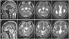
A 36-year-old woman visited a neurology department with a 7-day history of a throbbing headache and blurred vision in both eyes. Her medical history and physical examination were unremarkable. A neurological examination revealed hypoactive knee jerk, but all other results were normal. During the ophthalmologic examination, she complained of blurred vision but was able to count fingers at 2 meters. There was no diplopia or nystagmus. Her ocular movements, including the saccadic and smooth pursuit eye movements, were normal. There was no eyeball deviation or lid retraction. Both pupils were dilated (6 mm) and unresponsive to light. Light-near dissociation of the pupil was noted in both eyes, with bilateral tonic constriction to a near target followed by slow redilation. Both pupils were constricted (3 mm) after instilling topical 0.125% pilocarpine. The findings of laboratory tests, including cerebrospinal fluid and serologic tests for syphilis, were normal. The nerve conduction velocity and visual evoked potential were also normal. MRI showed dilation of the third ventricle and both lateral ventricles. There was no flow signal void at the aqueduct of Sylvius, which suggested the presence of obstructive hydrocephalus at that level (Fig. 1A). Ventricular-peritoneal shunting was performed 14 days after symptom onset (Fig. 1B). Four days after surgery, the size of the pupil and light reflex were completely restored in both eyes. Her symptoms did not recur during the 6-month follow-up.
Discussion
Adie's syndrome is a neurological disorder that affects the pupil of the eye and the autonomic nervous system. It presents with mydriasis, loss of deep tendon reflexes (DTRs), and diaphoresis. Ten percent of cases are bilateral and the prognosis is benign.1 Dorsal midbrain syndrome is characterized by several signs, including eyelid retraction, nystagmus, mid-dilated pupils with light-near dissociation, and paralysis of the upward gaze. It can be caused by obstructive hydrocephalus, multiple sclerosis, arteriovenous malformation, trauma or compression from a pineal tumor, or midbrain neurosarcoidosis.1 Our patient had early dorsal midbrain syndrome mimicking an Adie's tonic pupil, and cholinergic supersensitivity was demonstrated using topical 0.125% pilocarpine. Brain MRI revealed obstructive hydrocephalus at the level of the aqueduct of Sylvius, and her symptoms resolved 4 days after surgery.
Lesions in the Edinger-Westphal nucleus or anywhere along the course of the third nerve may damage the parasympathetic outflow and lead to pupillary dilation.6 Postciliary ganglionic lesions cause pupillary parasympathetic supersensitivity to a lower concentration of pilocarpine, but preciliary ganglionic lesions can also induce this condition.6 While a false-positive result is not unusual in a patient with cholinergic supersensitivity, and only 10% of tonic-pupil cases are bilateral, these findings suggested that Edinger-Westphal nuclear lesions can produce dorsal midbrain syndrome mimicking an Adie's tonic pupil. Most cases of tonic pupil are benign and do not warrant detailed investigations.1,2 However, the findings in our patient suggest that the early dorsal midbrain syndrome mimicking an Adie's tonic pupil was caused by obstructive hydrocephalus compressing the Edinger-Westphal nucleus.7
We do not have a plausible explanation for the transient tonic pupils and hypoactive knee jerk in our patient. DTRs may be markedly diminished or even absent in some patients even when there is no other evidence of nervous system disease. However, the primary problems in eliciting DTRs are the use of poor tools and poor technique.8
We believe that our case represented an early dorsal midbrain syndrome, with reversible Edinger-Westphal nucleus dysfunction and light-near dissociation. Physicians should remain vigilant for the possibility of early dorsal midbrain syndrome despite positive results on dilute-pilocarpine testing.
 XML Download