

 PDF
PDF ePub
ePub Citation
 Print
Print


Introduction
Muscular dystrophies are groups of inherited diseases of the muscle that are characterized clinically by progressive muscle weakness, and pathologically by muscle degeneration. Over the last 20 years, most of the genetic defects have been identified through the progress of genetics in this field. Even though there is currently no effective treatment for these diseases, gene therapy will provide the most promising answers to the problem. Duchenne muscular dystrophy (DMD) is the most common and most devastating form of muscular dystrophy, and so the search for treatment for these conditions has focused mainly on this disease.
Successful gene therapy and its development face difficulties; challenging factors include the sizes and complexities of the causative genes. However, new molecular strategies have been established to restore the genetic defect over the past few years (Table 1).
The present work reviews the theoretical background and recent progress in gene therapies for both DMD and other muscular dystrophies.
Gene Transfer
Plasmid vectors
Plasmid DNA is an extrachromosomal DNA molecule separated from chromosomal DNA. It is capable of replicating independently and has been used as a nonviral vector for gene transfer. Plasmid DNA can be engineered to include a dystrophin expression insert, and it can be expanded and purified in large amounts. When naked plasmid DNA containing a dystrophin cDNA construct was injected into the muscles of an mdx mouse (an animal model of DMD), dystrophin expression was observed in some of the muscle fibers.1 However, low efficiency was a limitation of a gene transfer system using a naked plasmid. To increase the efficacy of gene transfer, several physical manipulations were employed. Researchers demonstrated that the efficacy of gene transfer with intramuscular administration was much increased by the application of an electrical field across muscle fibers.2,3 Physical damage to the muscle induced by application of an electrical field was a major limitation of this approach, and subsequent research reports described measures to reduce this physical damage.4,5 One delivery method is in vivo electroporation, which reportedly increases the efficacy of gene transfer within muscles of an mdx mouse.6,7 In addition, the enzyme hyaluronidase, which can weaken the extracellular matrix of muscle fibers, could be used before electroporation, thus potentially further improving the efficacy of gene transfer and minimizing the physical damage to muscle fibers.8,9 However, it appears that a therapeutic application for this approach in DMD patients is unlikely.
Another method is a pressurized isolated-limb perfusion approach, which reportedly achieve a much higher efficiency than direct intramuscular injection in animal models.10 Zhang et al.11 temporarily blocked the circulation of a hindlimb in an mdx mouse with a preinjection of papaverine, and reperfused the limb with a large amount of plasmid DNA with a dystrophin gene insert. Cannulation of a large artery and the use of vasoactive agents were limitations of this approach. As an alternative, Hagstrom et al.12 demonstrated that a pressurized isolated-limb perfusion approach using a peripheral vein could also be effective in mammalian animal models. However, the transfection efficiency remained lower than that of viral vectors, even though it was markedly higher than that of intramuscular administration. Despite this, the capability of plasmid vectors to contain full-length dystrophin cDNA, as well as their noninfectious and nonimmunogenic properties, are major merits when compared to viral vectors.
In a phase I study, plasmid DNA containing a full-length dystrophin gene was injected into the radialis muscle in nine patients with either DMD or Becker muscular dystrophy (BMD).13 Neither serious adverse effects nor antidystrophin immune responses were found in the study. Dystrophin was expressed in six of the nine patients, although at a low level. A clinical trial of the delivery of plasmid DNA containing a full-length dystrophin gene using a high-pressure intravascular delivery approach is currently being conducted by the Transgene and Mirus consortium.
Viral vectors
Over recent years, adenovirus and adenoassociated virus (AAV) have been used as viral vectors of gene therapy for DMD. Adenovirus has double-stranded DNA (35 kb); adenoviral vectors inserted with the full coding sequence of dystrophin showed efficient and functional expression of dystrophin in the mdx mouse.14 However, an acute inflammatory response and immune reactions caused by their capsid proteins limit their use as the preferred viral vector for DMD.15
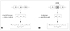
The AAV is a single-stranded DNA virus (4.7 kb) and requires a helper virus such as adenovirus or herpesvirus for its replication. The major advantages of an AAV vector are its mild immune response16 and high efficiency in transducing skeletal muscles than other vectors. However, its genome is too small for the insertion of a large dystrophin gene. To overcome this problem, dystrophin microgenes were produced by removal of most of the middle rod domain and portions of the amino- and carboxyl terminals of the dystrophin gene (Fig. 1). It was reported that minidystrophin and microdystrophin could improve the phenotype of muscular atrophy in an mdx mouse model.17,18 Intravenous delivery of the AAV also demonstrated widespread transduction of cardiac and skeletal muscles in an mdx mouse model.19
In limb-girdle muscular dystrophies (LGMD), gene transfer with AAV vectors was also investigated in LGMD2D (α-sarco-glycanopathy).20 In that research, human α-sarcoglycan (α-SG) was delivered with an AAV vector via local intramuscular injection in α-SG-knockout mice. Strong and persistent expression of the α-SG gene and restoration of the dystrophin-glycoprotein complex were observed without definite cytotoxicity.
The AAV vector is currently one of the most promising viral vectors; however, there remain additional problems to be solved. Although AAV vectors seem to induce a lower immune reaction compared to adenovirus, there has been evidence of an immune response against the myofibers transduced by these vectors.21 As a possible solution to this issue, Rivière et al.22 recently reported that the use of different serotypes for the subsequent injections of AAV vectors could sustain an efficient gene transfer in skeletal muscle. In addition, the titer of AAV in animal trials has been too high, so it is necessary to reduce the dose of virus to be applied to humans. One approach is to use AAV serotypes that are more capable of crossing the vascular endothelium. Recent studies have revealed increased efficiency of gene transfer to muscle fibers with the use of newly developed serotypes.23,24 Another way is the use of agents such as vascular endothelial growth factor, which can enhance the permeability of the microvasculature to increase the extravasation of the virus.19
A successful clinical trial of gene transfer of α-SG genes with an AAV vector was recently performed in patients with LGMD2D.25 That study was a double-blind, randomized controlled trial using an AAV vector containing α-SG genes injected locally in the extensor digitorum brevis muscle. Sustained expression of the α-SG gene and restoration of the dystrophin-glycoprotein complex were seen without any evidence of adverse events. A phase I/II clinical trial for DMD using an AAV vector for intramuscular delivery of microdystrophin into the biceps has been underway in the USA since 2006 (http://clinicaltrials.gov/ct2/show/NCT00428935, http://genetherapy.unc.edu/clinical.htm). AAV vectors are expected to be applied more to muscular dystrophies involving genes smaller than dystrophin.26
Exon Skipping Using Antisense Oligonucleotides
Deletion mutations in the dystrophin gene can induce either DMD or BMD, but the clinical phenotypes differ considerably. In 1988, Monaco et al.27 proposed a "reading-frame hypothesis". According to this hypothesis, the difference between these phenotypes is associated with a loss of frame (out-of-frame deletions) in DMD and a preservation of the reading frame (in-frame deletions) in BMD. About 65% of DMD cases are caused by out-of-frame deletions, which can produce truncated, nonfunctional dystrophins. On the other hand, in-frame deletions allow the expression of internally deleted but partially functional dystrophin proteins. Therefore, the DMD phenotype can be theoretically changed to the BMD phenotype by restoring the reading frames. The reading frame can be recovered by blocking a neighboring exon in out-of-frame deletions during pre-mRNA splicing (exon skipping, Fig. 2).28 Mann et al.29 reported a blocking of exon 23 by using 2-O-methyl antisense oligonucleotides (AONs) after local intramuscular injection in an mdx mouse. In their study, immunohistochemical staining verified the restoration of dystrophin and γ-SG in the subsarcolemmal area. To demonstrate the effectiveness of the systemic delivery, Alter et al.30 injected phosphorodiamidate morpholino (morpholino) AON into an mdx mouse to block exon 23. DMD mRNA of a size corresponding to the skipping of exon 23 was found by the reverse transcriptase-polymerase chain reaction in all skeletal muscles, and immunohistochemical staining revealed the restoration of dystrophin expression in skeletal muscles throughout the body, although the expression level differed considerably among muscles. Various kinds of AONs or antisense oligomers have been employed for this approach, including the phosphorodiamidate morpholino oligomers (PMOs), 2-O-methyl phosphorothioate (2OMe), and peptide-linked PMOs.29,31-33
The AON approach has some disadvantages. It needs regular repetitive administration, since the method modifies only the process of mRNA splicing. In addition, different AONs are needed for different gene deletions of dystrophin.
The clinical use of this approach was first reported by Takeshima et al.34 in 2006, when they intravenously injected phosphorothioate AON aiming at exon 19 into one DMD patient weekly for 4 weeks and observed the expression of dystrophin in a muscle biopsy. However, the clinical improvement was minimal. van Deutekom et al.35 recently reported the local effects of 2OMe AON aiming at exon 51 in DMD patients, and the results were quite promising. A single dose of 0.8 mg of AON was injected into the tibialis anterior muscle in four patients with DMD. Dystrophin expression was observed in 64-97% of myofibers along with specific skipping of exon 51 in muscle biopsies performed 28 days after injection. Adverse effects were not detected. Current phase I/II trials in the United Kingdom are monitoring the local intramuscular effects of a PMO aiming at exon 51 in children with DMD (http://clinicaltrials.gov/ct/gui/show/study/NCT00159250).
Readthrough of Stop-Codon Mutations
A small proportion of DMD patients (about 15%) exhibit nonsense mutations. Aminoglycoside antibiotics have been known to suppress stop-codon recognition. High-dose gentamicin therapy was reported to cause readthrough of premature stop codons and allow the restoration of dystrophin expression in mdx mice.36 However, two human trials of intravenous gentamicin have failed to show a definite benefit in patients with DMD and BMD.37,38
The readthrough efficiency of gentamicin was reported to vary markedly with different stop codons. The efficiency of translational readthrough is higher for UGA sequences than for UAG or UAA sequences. In addition, the nucleotide immediately downstream of the stop codon significantly influences the readthrough efficiency, in the order C>U>A≥G.39
Whilst two clinical trials have not found any toxicity associated with gentamicin, ototoxicity and nephrotoxicity are well-known adverse effects associated with its long-term use. As a result, searches for new drugs that do not exhibit these adverse effects but have better read-through efficiency have been initiated. One of those drugs, PTC124 (Ataluren, developed by PTC Therapeutics), can be taken orally and induces the read-through of premature stop codons. Welch et al.40 reported that PTC124 restores dystrophin production in primary muscle cells from DMD patients and mdx mice. It also improves the function of striated muscle in mdx mice within 2-8 weeks of drug administration and decreases serum creatine kinase levels.
The stop-codon readthrough approach also has some disadvantages. As described previously, the readthrough efficiency of gentamicin varies with different stop codons, and chronic administration is associated with adverse effects. However, the readthrough efficiency of PTC124 does not seem vary with different stop-codons.
A phase I trial found good tolerability of PTC124 in 62 healthy adult volunteers, with only some mild adverse effects, including dizziness, headache, and gastrointestinal disturbances, being noted.41 Phase II clinical trials of PTC124 in patients with DMD and BMD are in currently in progress at multinational centers (http://clinicaltrials.gov/ct2/show/NCT00264888, http://clinicaltrials.gov/ct2/show/NCT00592553).
Gene Modification for Cell Therapy
Cell therapy can be divided autologous or allogeneic. Even though genetic modification is not required in allogeneic cell therapy, the need for immunosuppression is a limitation. On the other hand, autologous cell therapy requires gene modification to restore the genetic defects. Quenneville et al.42 demonstrated the restoration of dystrophin by the cotransfection (nucleofection) of a plasmid vector containing the full-length human dystrophin gene and a phiC31 integrase in muscle precursor cells of mdx and severe combined immunodeficient (SCID) mice.
Lentivirus is currently one of the most promising viral vectors for an effective transfection of autologous cells due to its ability to incorporate into host chromosomes in various dividing cells. Lentiviral vectors expressing microdystrophin were reported to successfully transduce autologous satellite cells for transplantation into mdx mice.43 Nonmuscular-origin stem cells can also be used to restore dystrophin expression in muscles. Blood-derived CD133+ cells could be transduced with lentiviral vectors carrying a construct designed for axon skipping in dystrophic scid/mdx mice after intramuscular and intra-arterial delivery.44 In addition, Sampaolesi et al.45 demonstrated the expression of dystrophin by the transduction of mesoangioblasts with a lentiviral vector containing human microdystrophin in dystrophic dogs. Their study found that muscle function and motility were also improved, together with the restoration of dystrophin.
In a phase I clinical study, autologous muscle-derived CD 133+ stem cells were transplanted into eight patients with DMD. The muscle strength did not differ significantly between treated and untreated muscles across all patients, and no local or systemic adverse events were noted.46
Conclusion
Several promising strategies in gene therapy for muscular dystrophies that are currently being explored in clinical trials are reviewed herein. These strategies include gene transfer using nonviral and viral vectors, oligonucleotide-mediated exon skipping, stop-codon readthrough approaches, and gene modification for cell therapy. There remain some limitations and challenges to be resolved in all of these strategies. Within a few years the results of various clinical trials that are currently being undertaken will become available. It is highly anticipated that at least one or two of these strategies will prove efficacious and evolve into an effective gene therapy for patients with DMD, BMD, and other muscular dystrophies.


 XML Download