Journal List > J Clin Neurol > v.5(3) > 1048272

Yim, Ryu, Baik, Kim, Park, and Han: Common Carotid Artery Agenesis: Duplex Ultrasonographic Findings

Abstract

Background

Agenesis of the common carotid artery (CCA) resulting in separation of the origin of the external carotid artery (ECA) and internal carotid artery (ICA) from the aortic arch is rare. Fewer than 25 cases have been reported, and correlative ultrasound data were available for only 1 of them.

Case Report

A 52-year-old woman visited the hospital with a 3-day history of vertigo and headache. Color-coded duplex ultrasonography performed to evaluate the carotid and vertebral arteries revealed a normal configuration on the left side. However, the right CCA could not be found; instead, there were two vessels of approximately equal size in close proximity to each other. The cerebral angiographic findings were consistent with the ultrasonographic findings. The ECA and ICA originated directly from the brachiocephalic trunk, and the ECA arose proximal to the ICA.

Conclusions

The ultrasonographic findings revealed absence of the CCA, the ECA and ICA originating separately from the aortic arch. Color-coded duplex ultrasonography appears to be an effective and sensitive method for detecting absence of the CCA. These findings should help to further our understanding of the embryologic development of the carotid arteries.

Introduction

Agenesis of the common carotid artery (CCA) resulting in separation of the origin of the external carotid artery (ECA) and internal carotid artery (ICA) from the aortic arch is rare. Fewer than 25 cases have been reported, and correlative ultrasonographic data were available for only 1 of them.1-4 We report herein a case of absence of the CCA, the ECA and ICA originating separately from the aortic arch, as evidenced by color-coded duplex ultrasonography.

Case Report

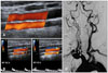
A 52-year-old woman with a history of hypertension and hypercholesterolemia visited the hospital with a 3-day history of vertigo and headache. The results of physical and neurological examinations were unremarkable. Color-coded duplex ultrasonography performed to evaluate the carotid and vertebral arteries revealed a normal configuration on the left side. No significant stenotic flow or plaque formation was observed in the left CCA, ECA, or ICA. However, the right CCA could not be found; instead, there were two vessels of approximately equal size in close proximity to each other. The more medially oriented vessel exhibited a low-resistance flow pattern consistent with the ICA, and the more laterally oriented vessel exhibited a high-resistance flow pattern consistent with the ECA (Fig. 1A-C). There was no significant stenotic flow or plaque formation in either of the two vessels. The cerebral angiographic findings were consistent with the ultrasonographic findings. The ECA and ICA originated directly from the brachiocephalic trunk, and the ECA arose proximal to the ICA (Fig. 1D).

Discussion

The embryologic development of this anomaly has been discussed previously by Lie.5 The CCA may be absent if the ductus caroticus (an embryologic vascular remnant) persists and there is involution of the distal portion of the third branchial arch. An alternative mechanism involves a failure of the ECA to migrate laterally and join the ICA (which arises from the third aortic arch) during development. As shown in this case, the resistive indices and spectral waveforms of the ICA and ECA are determined mainly by the regions and their supplied organs and not by the CCA.1,6 CCA agenesis is usually encountered during the diagnostic process. Color-coded duplex ultrasonography appears to be an effective and sensitive method for detecting absence of the CCA. In cases of carotid stenting, the technical problem of a lack of distal wire support in the ECA may lead to a higher risk of dislocation of the plaque and debris embolization in the ICA during the unprotected phase of the procedure.7 Endovascular treatment of these cases may be reserved for patients with stable plaques. We believe that the findings in our patient are instructive and will help to further our understanding of the embryologic development of the carotid arteries.

Figures and Tables

Fig. 1
Sonographic and angiographic findings of common carofid artery agenesis. (A) Color-coded duplex ultrasonography showing absence of the right common carotid artery (CCA). Instead, there are two vessels of approximately equal size in close proximity to each other. One vessel (B) shows a high-resistance flow pattern consistent with the external carotid artery (ECA), while the other vessel (C) shows a low-resistance flow pattern consistent with the internal carotid artery (ICA). (D) Cerebral angiography showing that the ICA (I, arrows) and ECA (E) originate directly from the brachiocephalic trunk, and that the ECA arises proximal to the ICA. The vertebral artery (V) is also shown.
jcn-5-149-g001

Acknowledgements

This work was supported by the 2006 Inje University research grant.

References

1. Woodruff WW 3rd, Strunsky VP, Brown NJ. Separate origins of the left internal and external carotid arteries directly from the aortic arch: duplex sonographic findings. J Ultrasound Med. 1995. 14:867–869.
crossref
2. Maybody M, Uszynski M, Morton E, Vitek JJ. Absence of common carotid artery: a rare vascular anomaly. AJNR Am J Neuroradiol. 2003. 24:711–713.
3. Simons D, Patetsios P, Moglia R, Dietz P. A case of congenital absence of the right common carotid artery: a rare embryologic anomaly. Journal for Vascular Ultrasound. 2003. 27:106–109.
crossref
4. Purkayastha S, Gupta AK, Varma DR, Bodhey NK, Vattoth S. Absence of the left common carotid artery with cervical origin of the right subclavian artery. AJNR Am J Neuroradiol. 2006. 27:708–711.
5. Lie TA. Congenital anomalies of the carotid arteries. 1968. Amsterdam: Excepta Medica Foundation.
6. Beach KW, Strandness DE Jr. Carotid artery velocity waveform ansVascular Disease. 1985. St. Louis: Mosby;410–413.
7. Faggioli GL, Testi G, Ferri M, Rossi C, Stella A. Common carotid agenesis and internal carotid stenting. Int Angiol. 2007. 26:290–291.
TOOLS
Similar articles