

 PDF
PDF ePub
ePub Citation
 Print
Print


Characteristic MRI findings of spontaneous intracranial hypotension or intracranial hypotension following lumbar puncture are pachymeningeal enhancement, subdural fluid collections or subdural hematoma, engorgement of cerebral venous sinuses, enlargement of the pituitary gland, crowding of the posterior fossa, and a decrease in the size of the prepontine cisterns.1,2 These abnormalities have been explained by the Monro-Kellie doctrine,3 whereby the summed volume of the brain, cerebrospinal fluid (CSF), and intracranial blood is nearly constant in an intact cranium. Therefore, the intracranial blood volume will be affected if CSF leakage occurs, resulting in a compensatory intracranial hyperemia that manifests as changes in brain MRI findings.
We unexpectedly found intracranial air in patients complaining of orthostatic headache. Intracranial hypotension with orthostatic headache is a rare cause of pneumocephalus,1,2 and here we report on these cases, including the imaging findings.
CASE REPORT
Case 1.
A 75-year-old man with congestive heart failure and atrial fibrillation was transferred to our hospital for the management of dyspnea and coughing. A chest x-ray indicated pulmonary edema. He had received posterolateral fusion with pedicle screw fixation of L4 and L5 foramina 5 years previously. However, he suffered from sustained painful dysesthesia on the right leg and received follow-up decompression surgery for L4 and L5 foramina stenosis 2 days before admission to our hospital, during which the dura was torn and dural plasty was performed.
On the 18th hospital day at the Department of Internal Medicine he complained of nuchal discomfort with headache. Neurological deficits were not observed and the headache was not aggravated by body position. After 3 days, a large egg-sized swelling on the wound site was detected without any symptoms or signs of infection. Opening of the skin and subcutaneous tissue to place a drain resulted in CSF flushing. A rubber drain was placed at the leakage site. After the procedure, severe headache developed when he raised his head, which was considerably relieved in the supine position although it did not completely disappear. He was referred to our neurology department.
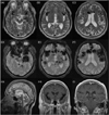
Neurological examination revealed no focal neurological deficits. Brain MRI with enhancement showed diffuse nonnodular pachymeningeal enhancement and massive pneumocephalus involving bilateral lateral ventricles, basal cisterns, sylvian cisterns, the left ambient cistern, retrocerebellar cisterns, cerebellar folia, the anterior interhemispheric fissure, and the subarachnoid space (Fig. 1). Emergency dural plasty was performed, and the headache disappeared.
Case 2.
A 54-year-old woman visited our hospital because of headache and vomiting without fever. Her headache was aggravated after sitting up and relieved immediately upon lying down. Two days before visiting our hospital, she received an epidural block in the sitting position by a midline approach for the management of back pain with radiating pain to her right leg. The air technique was applied to confirm that the needle was located in the epidural space, after which she felt that her head was being pulled to the ground for 10 minutes. Bupivacaine (0.5%) mixed with triamcinolone was injected at the level of L5-S1 after checking for the proper position of the needle in the epidural space. The next day she experienced the same headache twice while performing her daily routine. On the third day after the procedure, she suffered from severe orthostatic headache with vomiting and visited our hospital.
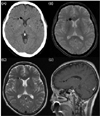
A neurological examination was normal. Brain CT showed a small amount of air in the right frontal horn. We performed brain MRI to evaluate the orthostatic headache (Fig. 2). Besides focal pneumocephalus, mild subdural hygroma in both anterior frontal areas with slightly prominent adjacent dural enhancement was observed. We also found more exaggerated pneumocephalus on gradient echo images. Her headache spontaneously regressed without complications. On follow-up brain CT performed 1 month later, mild subdural hygroma remained but the air had disappeared.
DISCUSSION
Pneumocephalus is defined as the presence of air within the cranial cavity and can occur after trauma, spinal surgery, craniofacial surgery, lumbar puncture, epidural steroid injections, gas-forming bacterial meningitis, Valsalva's maneuver, subarachnoid-pleural fistulae, and osteoma.1,4-10
In our first case, the 'inverted soda-bottle mechanism' could have been responsible for the pneumocephalus 4,5 - when a bottle containing soda is inverted, air rushes in to equilibrate the pressure differential as soda pours out. Similarly, as CSF flows out of the subarachnoid space through a dural arachnoid tear, a negative pressure is created within the subarachnoid space and the pressure differential results in air being drawn from the atmosphere to the lower pressure intracranial space. This air moves along CSF pathway - the subarachnoid space, cisterns (enlargements of subarachnoid space), and the ventricular system - via two foramina of Luschka and the foramen of Magendie.
In our second case there was no massive CSF leakage, so the injection of air while attempting to identify the epidural space via the loss-of-resistance-to-air technique might have been the cause of pneumocephalus, as reported in other cases.6,9-11 Such a loss-of-resistance-to-air technique is used to perform epidural block to locate the epidural space. During spinal tapping, resistance to the needle advancement after skin penetration is first encountered at the ligamentum flavum, and then the needle containing air arrives at the epidural space with a negative pressure (mean of - 7 cm of water), where air aspiration occurs. Since the dura is closely attached to the arachnoid, dural injury would rarely happen without damage to the arachnoid, and accidental dural puncture associated with the loss-of-resistance-to-air technique could cause pneumocephalus.
Whilst brain CT is a well-known diagnostic tool for detecting as little as 0.55 ml of intracranial air, gradient echo images are more sensitive. In fact, in our second case, brain CT was performed 1 day before brain MRI. Among brain CT, T2-weighted, and gradient echo MRI images in our patients, the last showed more-exaggerated pneumocephalus because of a paramagnetic effect of the air (Fig. 2). Therefore, in cases of orthostatic headache, it is preferable to perform gradient echo images in order to facilitate the detection of pneumocephalus.
Follow-up brain CT performed 1 month later revealed that the air had been absorbed but mild subdural fluid remained. Intracranial air tends to resolve spontaneously with rehydration, normalization of the CO2 level, N2O resorption, and replacement of CSF.5 Neither of our two patients showed cerebellar tonsillar descent, descent of the optic chiasm, reduction in the prepontine space, or crowding of the posterior fossa on brain images, instead showing pneumocephalus and good prognoses.
Kats et al. discriminated postdural puncture headache from pneumocephalus as follows: if the headache was relieved entirely by lying supine, it was identified as purely postdural puncture headache; however, if it was unchanged by position, it was diagnosed as purely resulting from intrathecal air.2 Other studies have similarly found that pneumocephalus caused a headache that was aggravated by any motion and was not relieved with lying down.1,9 However, our cases showed typical orthostatic headache despite the small degree of pneumocephalus, or more severe headache if a patient intended to raise his head, which was considerably relieved in the supine position.
We have been concerned about the existence of posterior fossa crowding or subdural hematoma in patients complaining of orthostatic headache, which indicates the need for surgical intervention or a poor outcome.12,13 When presented with cases of orthostatic headache, clinicians should keep in mind not only posterior fossa crowding and subdural hematoma but also pneumocephalus.
 XML Download