

 PDF
PDF ePub
ePub Citation
 Print
Print


INTRODUCTION
Stroke is one of major causes of death and disability in Western countries and in Korea. Due to the high mortality and morbidity rates associated with stroke, it is becoming a major community health problem worldwide. The association between heart disease and acute stroke is established.1 Cardiac sequelae, including myocytolysis, serum enzyme elevation, and arrhythmia, are known to develop in some ischemic stroke patients.2-6 The brain-heart connection was described early in the 20th century when Levy showed that changes in central nervous system metabolism influences cardiac function.7 Several studies have found that the levels of creatinine kinase.MB fraction (CKMB) and lactate dehydrogenase were elevated in 8-15% of patients with acute stroke,8,9 and that elevated levels of serum troponin T are found in 10-34% of patients with acute stroke.10-13 Serum cardiac troponin T (cTnT) is a new biochemical marker of myocardial damage with high specificity and sensitivity. There is considerable clinical and experimental evidence that cardiac changes in ischemic stroke result from excessive sympathetic nervous activity secondary to insular cortical damage.14 Several mechanisms can result in elevated concentrations of serum cTnT during the early phase of ischemic stroke, such as primary myocardial damage with secondary cardioembolic cerebral ischemia or primary cerebral ischemia, with secondary myocardial injury attributable to central activation of the sympathoadrenal system.9,15,16 Acute ischemic cerebrovascular events can induce various myocardial changes. Diffuse myocardial damage characterized by microislands of necrosis and subendocardial hemorrhage can occur after stroke.8,17-21 This type of injury is called myocytolysis, and it occurs as a result of intense activation of the sympathetic nervous system. A potential primary focus of myocytolysis is presumed to be its extensive autonomic and limbic connection.22,23
The aims of our study were to (1) identify the relationship between elevated serum cTnT and stroke severity, (2) determine the relationship between elevated serum cTnT and insular involvement, and (3) elucidate the influence of elevated serum cTnT on the prognosis associated with ischemic stroke.
MATERIALS AND METHODS
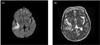
We identified 455 consecutive patients from a stroke database who were admitted to Kangbuk Samsung Hospital with a diagnosis of ischemic stroke between January 2005 and December 2006. The inclusion criteria were (1) acute cerebral infarction confirmed by brain diffusion-weighted imaging (DWI) magnetic resonance imaging (MRI) within 3 days of stoke onset; (2) measurement of serum cTnT level (≥0.01 ng/mL), and electrocardiography (ECG) and echocardiography performed within 3 days of stoke onset; and (3) a clear description of neurological scales. The exclusion criteria were (1) any recent ischemic heart disease, defined as previous myocardial infarction (AMI; American College of Cardiology/American Heart Association [ACC/AHA] criteria) within 2 weeks prior to and 3 days after stroke onset; (2) symptoms suggestive of AMI or unstable angina before admission; (3) newly developed pathologic Q waves on admission ECG; (4) previous coronary angioplasty or coronary bypass surgery; and (5) other heart diseases and debilitating diseases with the possibility of serum cTnT elevation, such as congestive heart failure, valvular heart disease (VHD), and end-stage renal disease (ESRD). The criteria for acute, evolving or recent AMI were defined by the ACC/AHA24 as the elevation of biochemical markers of myocardial necrosis (preferably troponin) with at least one of the following: (1) ischemic symptoms, (2) development of pathologic Q waves on the ECG, (3) ECG changes indicative of ischemia (ST segment elevation [≥1 mm] or depression [≥0.5 mm]), and (4) coronary artery intervention (e.g., coronary angioplasty). Serum cTnT was measured as a part of routine laboratory testing on admission to exclude subclinical coronary events. The presence of intracerebral or subarachnoid hemorrhage was ruled out by computed tomography at the time of admission. Thus, the study population comprised patients with acute ischemic stroke but without known or overt new ischemic heart disease. Information regarding heart disease symptoms, cardiac history, and risk factors for cardiovascular disease were obtained from the patients' medical records. Serum cTnT was measured from venous blood samples using the Elecsys 2010 platform (Roche Diagnostics, Mannheim, Germany). The cut-off value for elevated serum cTnT was set at 0.01 ng/mL. Twelve-lead ECG was performed immediately on admission. Of the original cohort, 39 patients were excluded for the following reasons: diagnosis of transient focal neurologic deficits (7 patients), cTnT measurement performed after 3 days of stroke onset (9 patients), cardiac problems and debilitating medical disease (16 patients with AMI, 3 with VHD, and 2 with ESRD), and incomplete medical records (2 patients). Blood pressures and pulse rates were measured immediately upon presentation at the emergency room. MRI was performed on a 1.5-T whole-body scanner (Intera Zyroscan; Philips Medical System, The Netherlands). Single-shot echo planar DWI imaging was performed with a repeat time of 7,500 ms, an echo time of 99.3 ms, a field of view of 22×22 cm, matrix of 128×128 cm, a slice thickness of 5 mm with a 1-mm gap, 23 axial slices, and b values of 0 s/mm2 and 1,000 s/mm2, with 6 gradient directions and 3 averages. Acute cerebral infarction was characterized by regions of increased signal intensity on DWI and decreased signal intensity on apparent diffusion coefficient maps. Acute infarctions on DWI were outlined manually on a slice-by-slice basis by a neuroradiologist and neurologist who were blinded to the clinical data. Insular involvement was defined as regions that involved the right or left insular cortex (posterior, superior, and medial areas; Fig. 1). To evaluate the clinical outcome in each patient, we applied a responder analysis that defined favorable outcomes at 30 days (and not 90 days) as influenced by the baseline National Institutes of Health Stroke Scale (NIHSS). Favorable outcome was defined using the following criteria: a 30-day modified Rankin Scale (mRS) score of 0 if the baseline NIHSS score was <8, an mRS score of 0 or 1 if the NIHSS score was 8-14, and an mRS score 0-2 if the NIHSS score was >14.25 Patients with an unfavorable outcome were defined as those not fulfilling the favorableoutcome criteria. The pathogenesis of stroke was classified by the Trial of Org 10172 in Acute Stroke Treatment (TOAST) classification.26,27
Categorical and continuous variables were compared by the chi-square test and Student's t-test, respectively. Univariate and multivariate logistic regression was used for further analysis. The cut-off for statistical significance was set at p value of 0.05. Analyses were performed with SPSS for Windows (version 12.0, Chicago, IL, USA).
RESULTS
A total of 416 patients with acute stroke, as confirmed by DWI within 72 h of stroke onset, were enrolled in this study. Serum cTnT was elevated in 10.8% (45/416) of the acute ischemic stroke patients. The 416 patients were divided into 2 groups according to serum cTnT levels: an elevated cTnT group (≥0.01 ng/mL) and a normal cTnT group (<0.01 ng/mL).
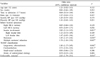
1. Comparison of the location and type of stroke between patients with elevated and normal serum cTnT
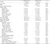
Epidemiologic and clinical characteristics were compared between the elevated and normal serum cTnT groups. Patients in the elevated serum cTnT group had a higher prevalence of atrial fibrillation and dyslipidemia than the normal serum cTnT group; they were also more likely to have multiple territorial infarctions and cardioembolism, but less likely to have deep penetrating artery infarction and, by TOAST classification, small vessel disease. Insular involvement was more common in the elevated serum cTnT group than in the normal serum cTnT group (56/371 [15.1%] vs. 13/45 [28.9%], p=0.042), but hemispheric laterality of insular involvement was not evident (Table 1).
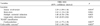
2. Comparison of severity and prognosis between patients with elevated and normal serum cTnT
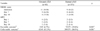
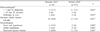
We compared stroke severity using the criterion of NIHSS score on the 1st day of admission. The stroke severity in the elevated serum cTnT group was greater and the disability was more severe (mRS on the 1st day) than in the normal serum cTnT group. The shortterm prognosis (30 days) was estimated by comparing the improvement using 30-day mRS responder analysis.25 The outcome was more favorable in the elevated serum cTnT group than in the normal serum cTnT group (3/45 [6.7%] vs. 72/371 [19.4%], p=0.015; Table 2). The univariate analysis showed that the elevated serum cTnT group was significantly associated with insular-lobe involvement, brainstem infarction, multiple territorial infarctions, cardioembolism and large-artery atherosclerosis, and unfavorable outcome (Table 3). Separate analysis of those variables identified in the univariate analysis for the independent association with elevated serum cTnT by multivariate analysis indicated that only insular-lobe involvement, cardioembolic stroke and unfavorable outcome were independently related to elevated serum cTnT (Table 4).
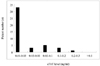
Levels of serum cTnT were less than 0.1 ng/mL in 91.1% of patients in the elevated cTnT group; they were more than 0.1 ng/mL in four (8.9%) of these patients, and the highest serum cTnT level in this group was 0.266 ng/mL (Fig. 2). Abnormalities on cardiologic evaluations including ECG, serum CK-MB, and echocardiogram were more frequent in the elevated serum cTnT group than in the normal serum cTnT group (Table 5).
DISCUSSION
Elevated serum cTnT was detected in 10.8% of the acute ischemic stroke patients that we examined, and was related to unfavorable outcomes 30 days after ischemic stroke. One of the novel findings of this study is that elevated serum cTnT in patients with ischemic stroke might be associated with stroke location and unfavorable outcome.
Every stroke is likely to exert considerable stress on the patient's heart. Elevated serum cTnT might be indicative of a lower cardiac tolerance to stress caused by the acute ischemic stroke. This is possible explanation for the relationship between elevated serum cTnT and the poor short-term prognosis found in this study. Previous studies have suggested that cardiac damage after ischemic stroke is associated with sympathoadrenal activation.14,20 Epinephrine and cortisol concentrations are elevated after a stroke and higher concentrations have been reported in association with myocardial damage.10 In the present study, elevated serum cTnT level was associated with severe stroke and insular-lobe damage, regardless of laterality, which can cause severe stress and lead to sympathoadrenal activation.
This study demonstrates that there is a relationship between elevated serum cTnT and stroke with insular cortex involvement. Sensory, autonomic, and limbic functions are integrated in the insula through reciprocal connections with principle sensory areas, paralimbic areas in the orbital, temporopolar and cingulate cortices, and the hypothalamus. Stimulation of the insula in rats as well as in humans undergoing surgery for epilepsy has demonstrated that this brain area is involved in cardiac autonomic control.13,28,29 The short-term outcome is less favorable and the stroke is more severe in stroke patients with elevated serum cTnT than in those with normal serum cTnT levels. Cardioembolic stroke was more common in patients with elevated serum cTnT. Doctors in charge of stroke units should be more prudent when dealing with these patients and should offer cardiologic evaluations and secondary prevention measures, and also investigations for stroke risk factors and proper interventions for coronary artery disease. When excluding patients with obvious cardiac damage from the study population, patients in the elevated serum cTnT group (0.16±0.15 µg/L, mean±SD) did not satisfy the clinical, ECG or echocardiographic criteria for AMI (ACC/AHA criteria). The ST segment elevations and depressions were not prominent enough to meet the criteria for AMI as defined by the ACC/AHA. Two of patients had developed pathologic Q waves since their previous ECG, which had been taken more than 6 months before that performed for the current study. None of the patients in the elevated serum cTnT group had chest pain. These findings might be attributable to serum cTnT being a more sensitive for indicator of myocardial damage. However, the elevation of serum cTnT might be related to underlying coronary artery disease exacerbated by the stress of an acute ischemic stroke. Our study was subject to several limitations. First, we relied on single baseline blood samples and did not retest serum cTnT so as to determine trends in the cTnT level, and thus we were unable to account for variations in serum cTnT levels that might have occurred over time. Repeated assays could provide additional information on the development and evolution of myocardial damage in patients with acute ischemic stroke. Second, we did not measure the concentrations of epinephrine, norepinephrine and cortisol as indirect markers of sympathoadrenal activation, which might have provided further information on the tone of the sympathetic nervous system. Third, our study was unable to assess the long-term prognosis in normal and elevated serum cTnT groups due to the short follow-up period. Despite these limitations, the findings of this study will improve the understanding of the implications of serum cTnT elevation in acute ischemic stroke. We suggest that clinicians should consider adding a serum cTnT titer to their routine admission testing in patients with suspected ischemic stroke, since a high serum cTnT level appears to be related to a significantly higher risk for worse outcomes and cardiac complications. Information on serum cTnT might also provide clues as to the location and extent of ischemic stroke.
In summary, elevated serum cTnT concentration without evidence of myocardial lesion was found in 10.8% of the acute ischemic stroke patients that we examined. Stroke severity (as assessed by the NIHSS and mRS) and insular-lobe involvement were associated with increased serum cTnT levels. Patients with elevated serum cTnT levels during acute ischemic stroke treatment showed worse outcomes than those with normal serum cTnT levels. Serial serum cTnT assessment and long-term clinical outcome data in a larger study population are needed in future studies in order to clarify the clinical implications of elevated serum cTnT levels in acute ischemic stroke.
 XML Download