

 PDF
PDF ePub
ePub Citation
 Print
Print


Fahr's disease is characterized by idiopathic calcification of the basal ganglia, dentate nuclei of the cerebellum, and the centrum semiovale,1,2 and is occasionally associated with either hypoparathyroidism or pseudohypoparathyroidism.1 Both familial and nonfamilial forms have been reported, with autosomal-dominant inheritance predominating.3-5 Clinically, it may present with movement disorders such as rigidity, hypokinesia, tremor, choreoathetosis, and ataxia, as well as behavioral disturbances such as dementia and mood disorders.5-9
Whilst parkinsonism is a well-recognized symptom associated with striopallidodentate calcification, prominent postural instability with frequent falling down and supranuclear gaze palsy, which are the characteristic clinical manifestations of progressive supranuclear palsy (PSP), have been very rarely reported.10-12
We describe a patient with bilateral striopallidodentate calcification who presented with progressive gait disturbance, prominent postural instability, oculomotor disturbances, and dementia.
CASE REPORT
A 70-year-old woman was admitted to hospital because of bilateral pleural effusion, and referred to our neurology department due to a 2-year history of gait disturbance that occasionally resulted in falling down, before which she had been well. Six months after the first episode of falling down, her family noticed that she had a masked face, monotonous speech, and bradykinesia in both arms and legs. During the subsequent year she fell down more frequently, and she could not walk without assistance. She had been taking vitamin D and a calcium preparation to treat a 10-year history of idiopathic hypoparathyroidism and recurrent compression fracture of the thoracic and lumbar vertebral bodies. There was no family history of parkinsonism or any neurological disorders.
On examination, she was oriented to place and people, but had difficulty copying, calculating, and maintaining attention. A neuropsychological investigation showed a severe impairment in memory function, reduced psychomotor speed, and visuospatial dysfunction (Table 1). She had a decreased blink rate, a paucity of movement of the face, and mild dysarthria. Horizontal voluntary saccades were slightly limited and hypometric, and vertical upward and downward saccades were extremely limited and slow. Similarly, pursuit movements were full horizontally, but restricted on vertical gaze. She had a full range of eye movement in the doll's head maneuver, demonstrating that her gaze palsy was caused by a supranuclear problem. Other cranial nerve functions were normal. All four limbs exhibited bradykinesia and rigidity. Sitting up in a bed resulted in spontaneous retropulsion. She stood on a narrow base, and her stride was reduced with decreased arm swing bilaterally. She showed postural instability, and mild hesitation when initiating gait. The deep tendon reflexes were normoactive in all four limbs. However, there was no dystonia, pyramidal signs, or autonomic impairment.
Laboratory studies at admission showed the following values: 6.6 mg/dL serum total calcium (normal: 8.6 to 10.0 mg/dL), 4.99 mg/dL phosphate (normal: 2.7 to 4.5 mg/dL), 52 U/L alkaline phosphatase (normal: 25 to 100 U/L), and 1.44 pg/mL intact-parathyroid hormone (normal: 13.0 to 54.0 pg/mL). Serum thyroxin and thyroid-stimulating hormone levels were normal. The electrocardiogram showed a prolonged QTc interval of 0.56 seconds.
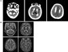
Brain computed tomography (CT) demonstrated extensive, bilateral calcification of the basal ganglia, centrum semiovale, and bilateral dentate nuclei of the cerebellum (Fig. 1-A). On magnetic resonance imaging there were mixed high-and low-intensity signals compatible with the distribution of calcification on CT. The red nucleus and substantia nigra appeared normal (Fig. 1-B). Single-photon-emission computed tomography using 99mTc-hexamethylpropylene amineoxime showed no abnormal perfusion defects in the brain.
Levodopa was started at 100 mg twice daily, and the dosage was slowly increased to 1000 mg per day. However, this did not produce any clinical improvement.
DISCUSSION
We experienced a case of striopallidodentate calcification associated with idiopathic hypoparathyroidism presenting with prominent oculomotor disturbances. The patient also showed parkinsonism that did not respond to levodopa treatment. The prominent oculomotor disturbances, in combination with these extrapyramidal features and dementia, suggested a clinical diagnosis of PSP.13
The origin of the parkinsonian symptoms and oculomotor disturbances of this patient is difficult to establish. One possibility is that the basal ganglia calcification observed in brain CT was an incidental finding, because asymptomatic basal ganglia calcifications may be seen at autopsy, especially in the elderly.14 However, the probability of such a coincidence is extremely low given the reported prevalence.15,16 In addition, it has been suggested that a metabolic disturbance such as low calcium or a high phosphorous plasma level may cause dopaminergic dysfunction in the substantia nigra pars compacta.17
18F-deoxyglucose and positron-emission tomography have shown that glucose metabolism in the basal ganglia and the frontal brain is massively reduced in Fahr's disease.9,18 This abnormality possibly results from a disruption of frontostriatal circuits, presumably at the level of the basal ganglia.9 The precise mechanism of oculomotor disturbance in our patient was unknown. In view of the co-occurrence of oculomotor abnormalities in other progressive basal ganglia disorders such as Parkinson's disease, PSP, and Huntington's disease, the significant impairment of eye movement in our patient may have been attributable to dysfunction of the basal ganglia in the generation of voluntary saccades.19
In conclusion, we have presented a rare case with idiopathic hypoparathyroidism complicated by oculomotor disturbances and parkinsonism with a PSP-like phenotype.

 XML Download